. 2018 Dec;16(6):4927-4942.
doi: 10.3892/etm.2018.6831. Epub 2018 Oct 5.

Hypothermic treatment after computer-controlled compression in minipig: A preliminary report on the effect of epidural vs. direct spinal cord cooling

Affiliations

Hypothermic treatment after computer-controlled compression in minipig: A preliminary report on the effect of epidural vs. direct spinal cord cooling

Monika Zavodska et al. Exp Ther Med. 2018 Dec.

Abstract

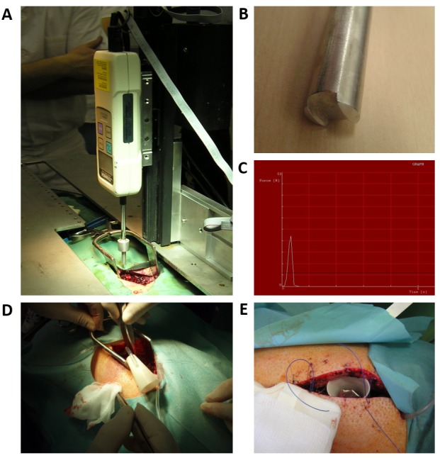
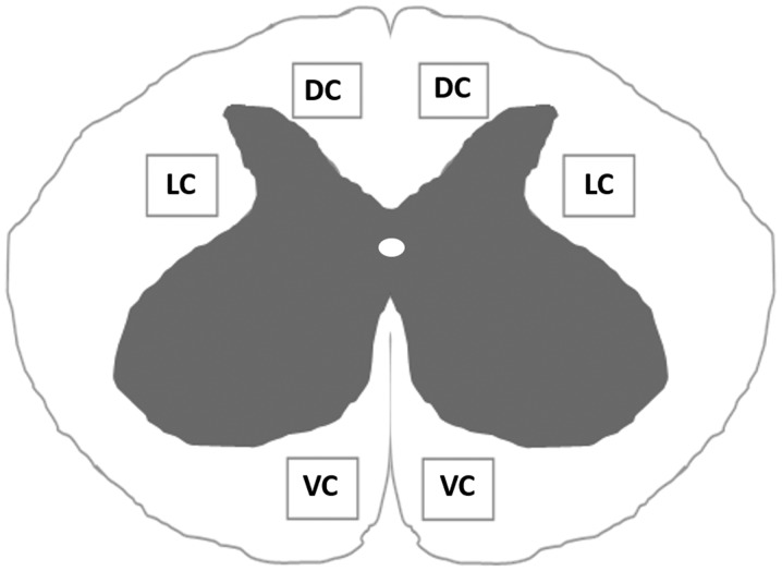
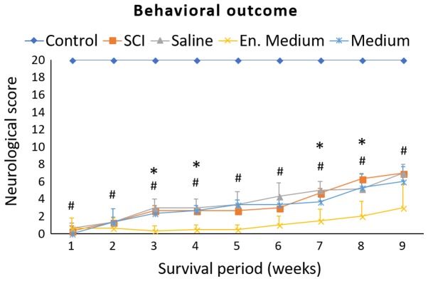
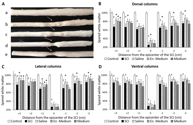
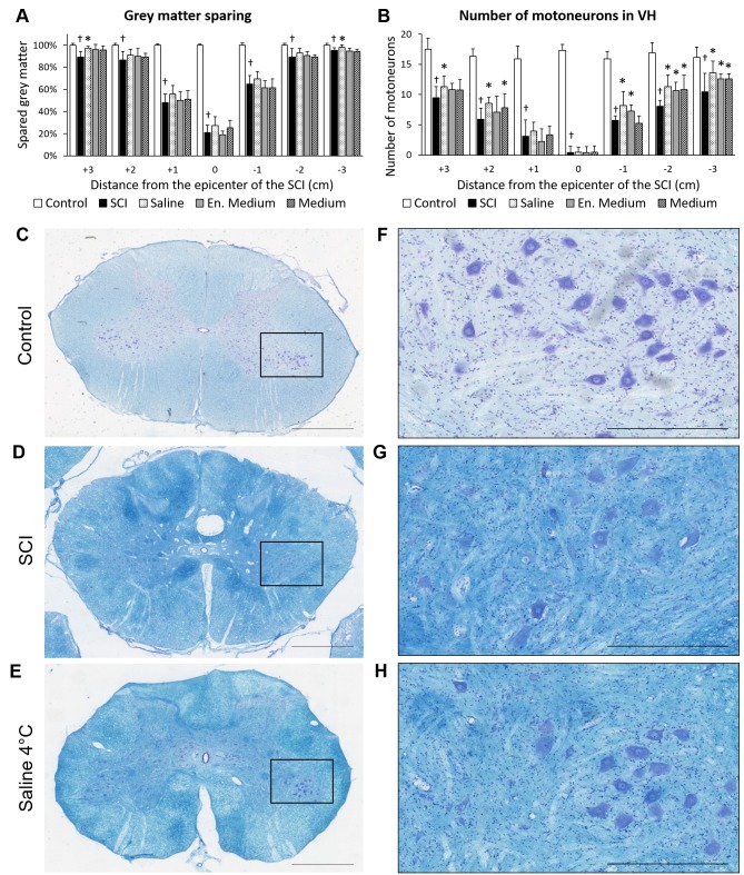
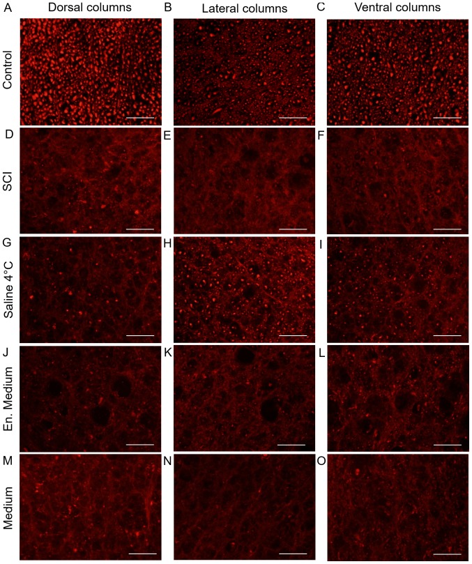
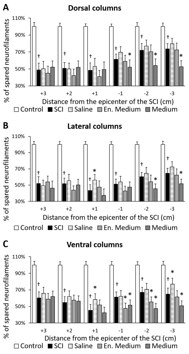
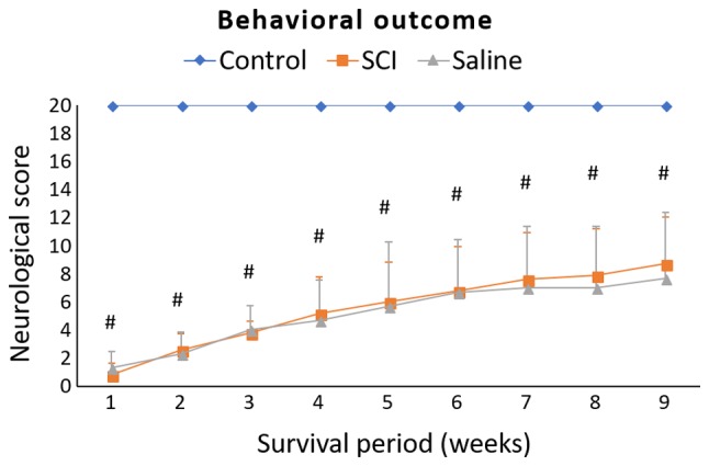
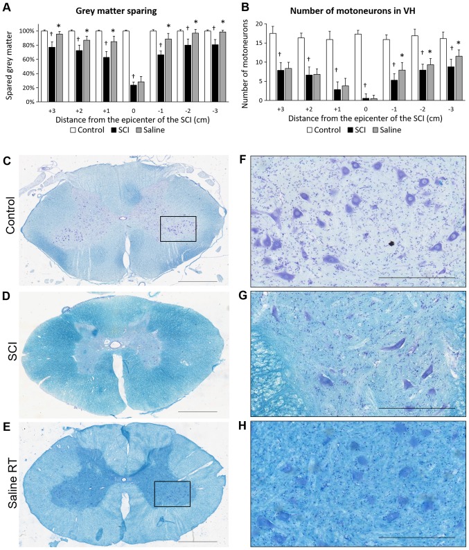
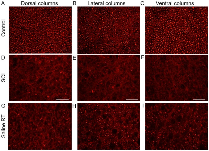
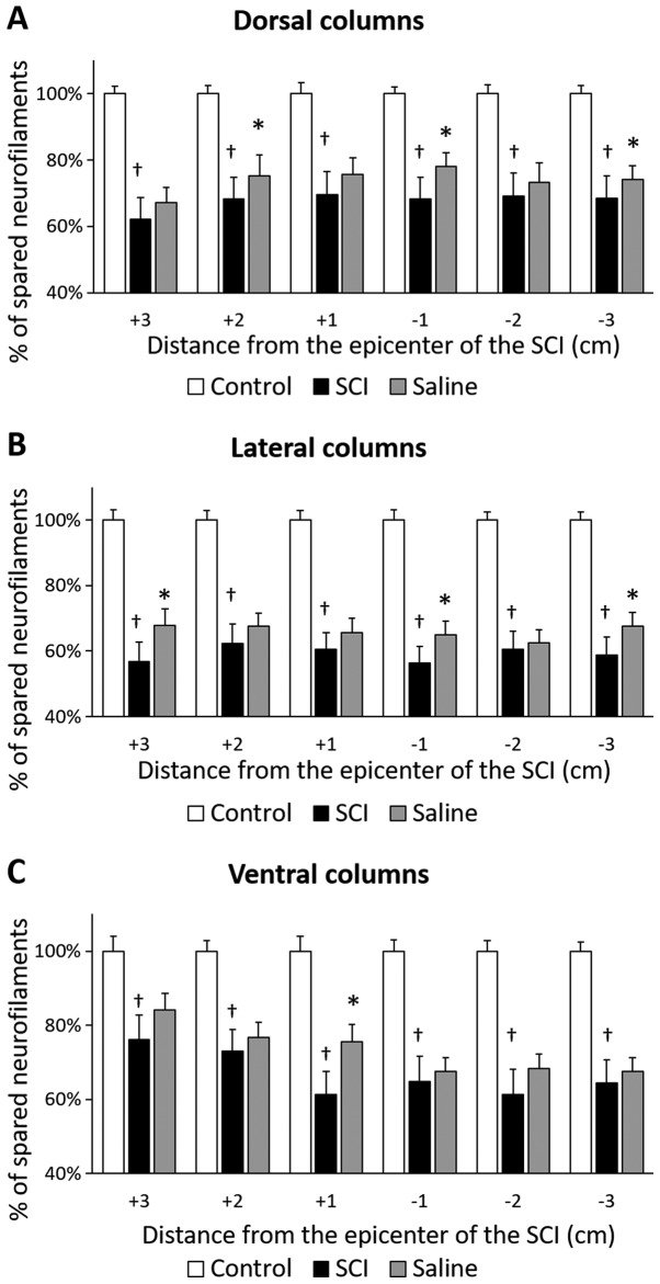
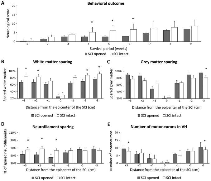
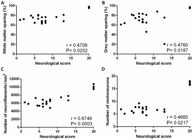
The aim of the present study was to investigate the therapeutic efficacy of local hypothermia (beginning 30 min post-injury persisting for 5 h) on tissue preservation along the rostro-caudal axis of the spinal cord (3 cm cranially and caudally from the lesion site), and the prevention of injury-induced functional loss in a newly developed computer-controlled compression model in minipig (force of impact 18N at L3 level), which mimics severe spinal cord injury (SCI). Minipigs underwent SCI with two post-injury modifications (durotomy vs. intact dura mater) followed by hypothermia through a perfusion chamber with cold (epidural t≈15°C) saline, DMEM/F12 or enriched DMEM/F12 (SCI/durotomy group) and with room temperature (t≈24°C) saline (SCI-only group). Minipigs treated with post-SCI durotomy demonstrated slower development of spontaneous neurological improvement at the early postinjury time points, although the outcome at 9 weeks of survival did not differ significantly between the two SCI groups. Hypothermia with saline (t≈15°C) applied after SCI-durotomy improved white matter integrity in the dorsal and lateral columns in almost all rostro-caudal segments, whereas treatment with medium/enriched medium affected white matter integrity only in the rostral segments. Furthermore, regeneration of neurofilaments in the spinal cord after SCI-durotomy and hypothermic treatments indicated an important role of local saline hypothermia in the functional outcome. Although saline hypothermia (24°C) in the SCI-only group exhibited a profound histological outcome (regarding the gray and white matter integrity and the number of motoneurons) and neurofilament protection in general, none of the tested treatments resulted in significant improvement of neurological status. The findings suggest that clinically-proven medical treatments for SCI combined with early 5 h-long saline hypothermia treatment without opening the dural sac could be more beneficial for tissue preservation and neurological outcome compared with hypothermia applied after durotomy.

Keywords: durotomy; local hypothermia; motoneurons; neurofilaments; spinal cord; white matter integrity.


Figures

Figure 1.
Figure 1.
(A) Computer-controlled apparatus for induction of contusive SCI. (B) Stainless steel rod with 5 mm diameter causing SCI when it impacts on the spinal cord. (C) Real impact force recorded during SCI. (D and E) Perfusion chamber implanted above the epicenter of SCI for inducing local hypothermia. SCI, spinal cord injury.
Figure 2.
Figure 2.
Schematic illustration of regions of interest. The number of neurofilaments was quantified in the DC, LC and VC. Measuring frame size in each area was 800×600 µm. DC, dorsal columns; LC, lateral columns; VC, ventral columns.
Figure 3.
Figure 3.
Experiment 1. Behavioral recovery of animals during 9-week survival after SCI and subsequent hypothermia with cold saline, en. medium or medium. The scoring points range from complete paraplegia (point zero) to normal hindlimb function (point 20). Data are the mean values ± standard deviation. The results were statistically evaluated for each week separately using one-way analysis of variance followed by Sidak's multiple comparison test. #P<0.0001, control vs. SCI; *P<0.05, SCI vs. hypothermia with en. medium. SCI, spinal cord injury; en. medium, enriched medium.
Figure 4.
Figure 4.
Experiment 1. (A) Macroscopic images of spinal cord and white matter sparing in spinal cord columns after SCI and hypothermic treatments. Spinal cords of animals in the (Aa) control, (Ab) 9 weeks after SCI, (Ac) after subsequent hypothermia with cold saline, (Ad) enriched medium, or (Ae) medium were indicated. Arrows demonstrated the epicenter of injury. White matter sparing after SCI and hypothermic treatment in (B) dorsal, (C) lateral and (D) ventral columns. Data are the mean values ± standard deviation. Ordinary one-way analysis of variance followed by Sidak's multiple comparison test was used for the assessment of differences between experimental groups in each segment separately. P<0.0001, vs. Control; *P<0.05 vs. SCI. SCI, spinal cord injury; en. medium, enriched medium.
Figure 5.
Figure 5.
Experiment 1. (A and B) Gray matter sparing and preservation of motoneurons 9 weeks after SCI and hypothermic treatment with saline, en. medium or medium, performed on transverse sections dyed with Luxol fast blue and cresyl violet staining. Transverse spinal cord sections taken from caudal segments (−3) of (C) control, (D) after SCI and (E) treatment with cold saline. (F-H) High-power microphotographs of the boxed areas in C-E indicated motoneurons. Data are demonstrated as the mean ± standard deviation. Ordinary one-way analysis of variance followed by Sidak's multiple comparison test was used for the assessment of differences between experimental groups in each segment separately. (A) Grey matter sparing. P<0.001 vs. Control; *P<0.05, vs. SCI. (B) Number of motor neurons in VHs. P<0.0001 vs. Control; *P<0.05 vs. SCI. Scale bar in C-E, 2 mm; in F-H, 500 µm. SCI, spinal cord injury; en. medium, enriched medium; VH, ventral horn.
Figure 6.
Figure 6.
Experiment 1. Representative images of neurofilament immunohistochemistry. Sections were taken from +1 segment of transverse spinal cord sections (dorsal, lateral and ventral columns) from (A-C) control, (D-F) after SCI and subsequent hypothermic treatment with (G-I) cold saline, (J-L) en. medium or (M-O) medium. Scale bar, 100 µm. SCI, spinal cord injury; en. medium, enriched medium.
Figure 7.
Figure 7.
Experiment 1. Number of neurofilaments (expressed as percentage of NF/mm2) in the (A) dorsal, (B) lateral and (C) ventral columns 9 weeks after SCI and hypothermic treatment with cold saline, en. medium or medium. The number of neurofilaments was evaluated bilaterally from ten randomly-selected transverse slices of each spinal cord block (3 cm rostrally and 3 cm caudally from the epicenter of injury). Data are presented as the mean ± standard deviation. Ordinary one-way analysis of variance followed by Sidak's multiple comparison test was used for the assessment of differences between experimental groups; each segment was assessed separately. P<0.0001 vs. Control; *P<0.05 vs. SCI. SCI, spinal cord injury; en. Medium, enriched medium.
Figure 8.
Figure 8.
Experiment 2. Behavioral recovery of animals during 9-week survival period after SCI and subsequent hypothermia with saline. The scoring points range from complete paraplegia (point zero) to normal hindlimb function (point 20). Data are presented as the mean ± standard deviation. Ordinary one-way analysis of variance followed by Sidak's multiple comparison test was used for the statistical analysis and each week was assessed separately. #P<0.0001, Control vs. SCI. SCI, spinal cord injury.
Figure 9.
Figure 9.
Experiment 2. (A) Macroscopic images of spinal cords and white matter sparing in spinal cord columns 9 weeks after SCI and saline treatment. Arrows point to the epicenter of the injury. Spinal cord of (Aa) control, (Ab) after spinal cord injury and (Ac) subsequent hypothermic treatment with saline at room temperature. White matter sparing in (B) dorsal, (C) lateral and (D) ventral columns. Data are the mean values ± standard deviation. Ordinary one-way analysis of variance followed by Sidak's multiple comparison test was used for the assessment of differences between experimental groups in each segment separately. P<0.0001 vs. Control; *P<0.05 vs. SCI. SCI, spinal cord injury.
Figure 10.
Figure 10.
Experiment 2. Gray matter sparing and preservation of motoneurons 9 weeks after SCI and hypothermic treatment with saline performed on transverse sections dyed with Luxol fast blue and cresyl violet staining (A-B). Transverse spinal cord sections taken from caudal segments (−3) of (C) control, (D) after SCI and (E) treatment with saline. (F-H) High-power microphotographs of the boxed areas in C-E showing motoneurons. Data are the mean values ± standard deviation. Ordinary one-way analysis of variance followed by Sidak's multiple comparison test was used for the assessment of differences between experimental groups in each segment of spinal cord separately. (A) Grey matter sparing; P<0.05 vs. Control; *P<0.05 vs. SCI. (B) Number of motor neurons in VHs; P<0.0001, vs. Control; *P<0.05 vs. SCI. Scale bar in C-E, 2 mm; in F-H, 500 µm. SCI, spinal cord injury; VH, ventral horn.
Figure 12.
Figure 12.
Experiment 2. Number of neurofilaments (expressed as percentage of neurofilaments/mm2) in white matter columns 9 weeks after SCI in the (A) dorsal, (B) lateral and (C) ventral columns after hypothermic treatment with saline. The number of neurofilaments was evaluated bilaterally from ten randomly-selected transverse slices of each spinal cord block (3 cm rostrally and 3 cm caudally from the epicenter of injury). Data are presented as the mean ± standard deviation. Ordinary one-way analysis of variance followed by Sidak's multiple comparison test was used for the assessment of differences between experimental groups in each segment separately. P<0.0001 vs. Control; *P<0.05 vs. SCI. SCI, spinal cord injury.
Figure 11.
Figure 11.
Experiment 2. Representative images of neurofilament immunohistochemistry. Sections were taken from +1 segment of transverse spinal cord sections (dorsal, lateral and ventral columns) from (A-C) control, (D-F) after SCI and subsequent (G-I) hypothermic treatment with saline. Scale bar, 100 µm. SCI, spinal cord injury; RT, room temperature.
Figure 13.
Figure 13.
Comparison of outcome parameters in SCI groups (SCI opened and SCI intact dura mater). The animals survived for 9 weeks. Data are the mean values ± standard deviation. Unpaired Student t test was used for statistical evaluation of data. *P<0.05 SCI opened vs. SCI intact. (A) Behavioral recovery, (B) cumulative white matter sparing, (C) cumulative grey matter sparing, (D) number of neurofilaments and (E) number of motoneurons in VHs were assessed. SCI, spinal cord injury; VH, ventral horn.
Figure 14.
Figure 14.
Scatterplots illustrating relationship between final neurological score of animals from all experimental groups and (A) cumulative white matter sparing, (B) cumulative grey matter sparing, (C) number of neurofilaments and (D) number of motoneurons in spinal cord VHs. Indicated are the Spearman rank correlation coefficients (r) and the P-values. The number of XY pairs is 24. VH, ventral horn.

Similar articles

Cited by

References

    1. Tator CH. Review of treatment trials in human spinal cord injury: Issues, difficulties, and recommendations. Neurosurgery. 2006;59:957–987. doi: 10.1227/01.NEU.0000245591.16087.89. - DOI - PubMed
    1. Tator CH. Update on the pathophysiology and pathology of acute spinal cord injury. Brain Pathol. 1995;5:407–413. doi: 10.1111/j.1750-3639.1995.tb00619.x. - DOI - PubMed
    1. McDonald JW, Sadowsky C. Spinal-cord injury. Lancet. 2002;359:417–425. doi: 10.1016/S0140-6736(02)07603-1. - DOI - PubMed
    1. Balentine JD. Pathology of experimental spinal cord trauma. I. The necrotic lesion as a function of vascular injury. Lab Invest. 1978;39:236–253. - PubMed
    1. Simard JM, Woo SK, Aarabi B, Gerzanich V. The Sur1-Trpm4 channel in spinal cord injury. J Spine Suppl. 2013;4:002. - PMC - PubMed