. 2018 Dec;16(6):5286-5297.
doi: 10.3892/etm.2018.6846. Epub 2018 Oct 11.

Atorvastatin reverses the dysfunction of human umbilical vein endothelial cells induced by angiotensin II

Affiliations

Atorvastatin reverses the dysfunction of human umbilical vein endothelial cells induced by angiotensin II

Haiming Dang et al. Exp Ther Med. 2018 Dec.

Abstract

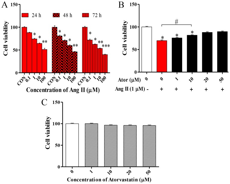
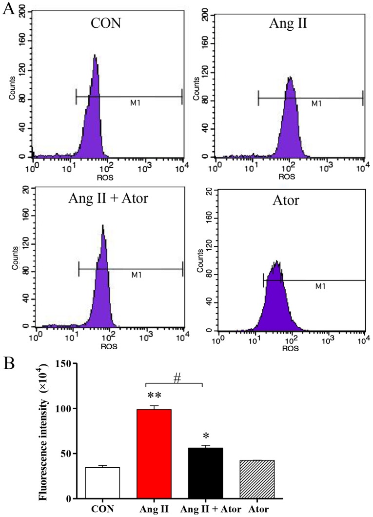
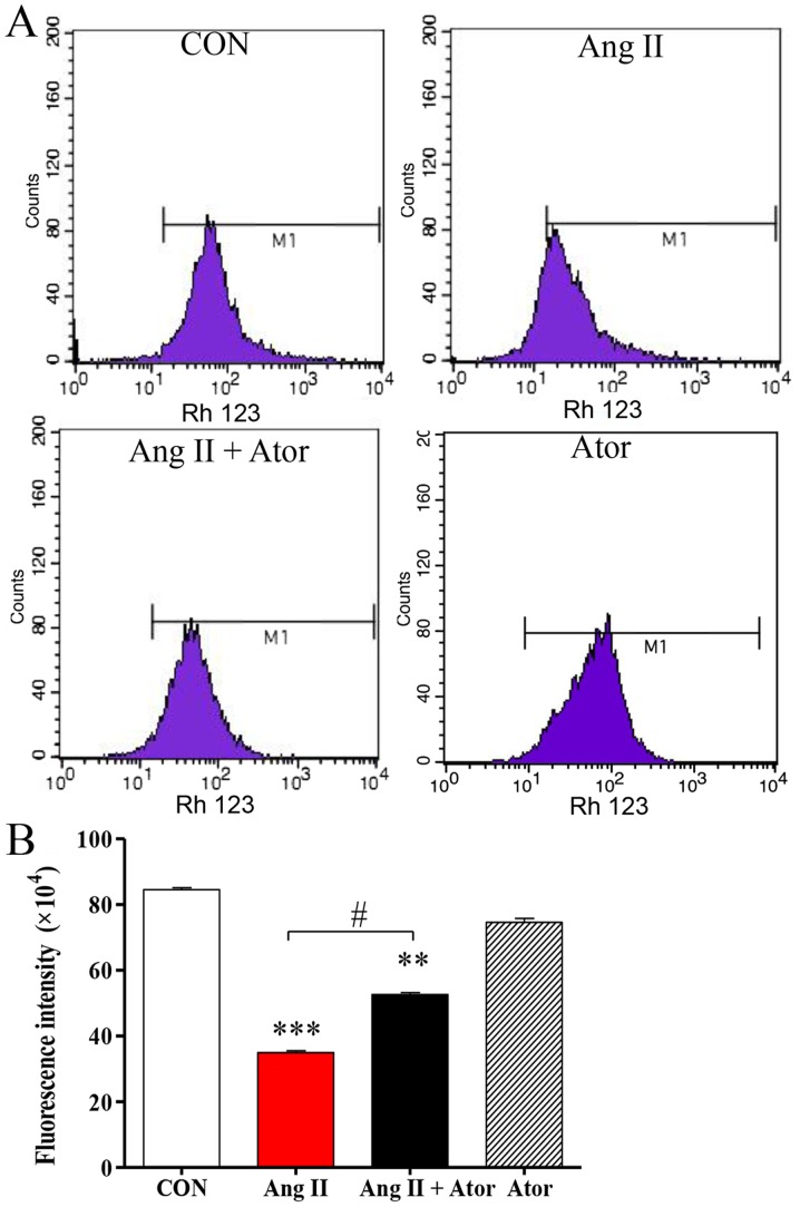
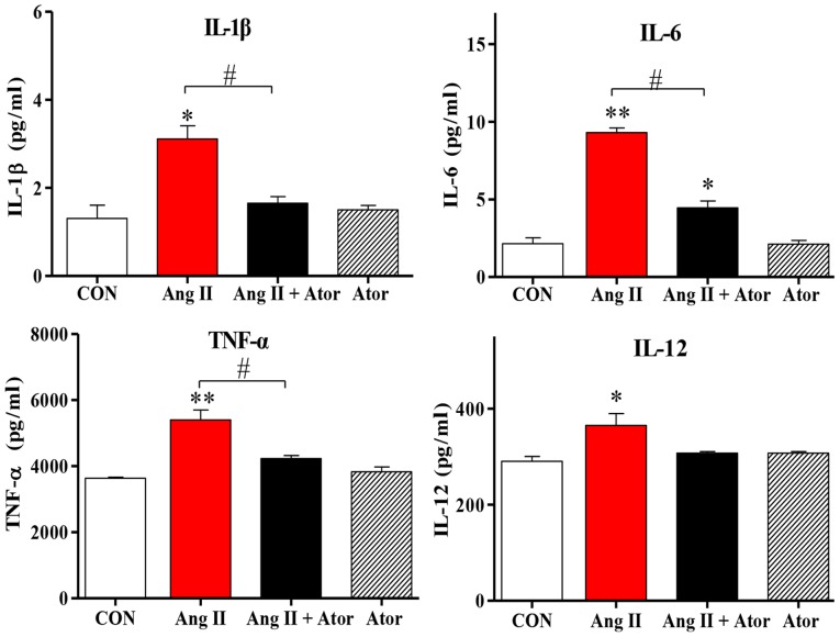
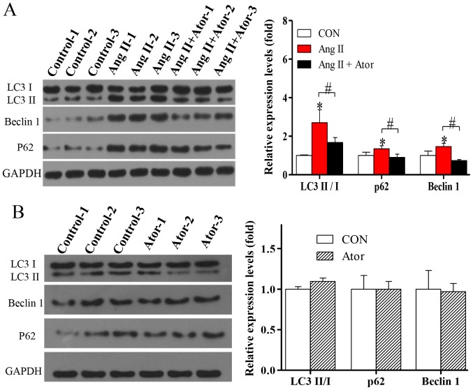
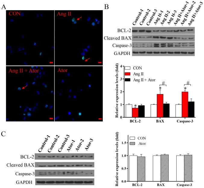
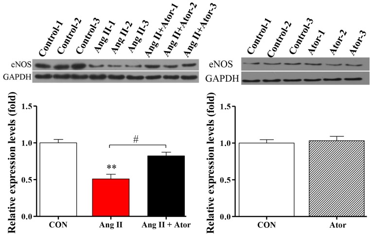
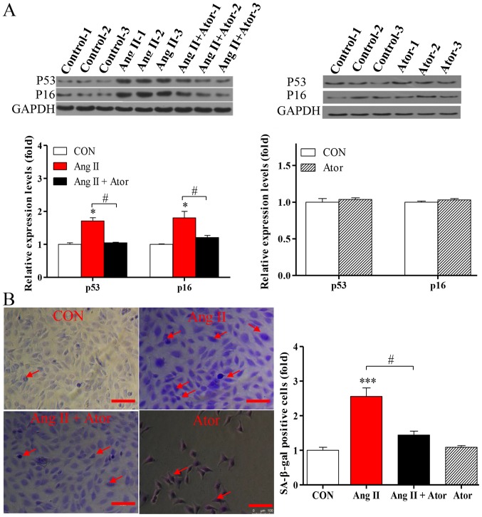
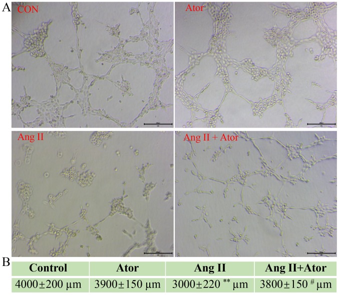
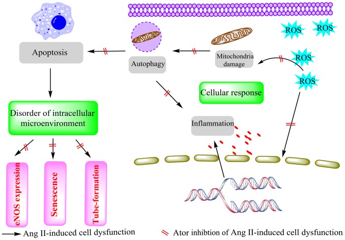
Statins exert pleiotropic effects on endothelial cells, in addition to lowering cholesterol. This study evaluated angiotensin II (Ang II)-induced dysfunction in human umbilical vein endothelial cells (HUVECs), and the effects of atorvastatin (Ator) on induced HUVECs in vitro. The cytotoxicity of Ang II and Ator was determined by the MTT assay. A series of cellular responses were screened, including oxidative stress, cellular apoptosis, inflammatory response, autophagy, expression of endothelial nitric oxide synthase and the angiogenic function of HUVECs. Ator returned these cellular responses to a normal level. The present study also examined cellular organelle dysfunction. In HUVECs, Ang II triggered mitochondrial damage, as demonstrated by a decreased mitochondrial membrane potential, while Ator attenuated this Ang II-induced damage. The observed cellular dysfunction may cause endothelial senescence due to excessive cell injury. The current study examined several aging markers, which revealed that these disorders of cellular functions triggered endothelial senescence, which was delayed by Ator. Ator also suppressed Ang II-induced angiogenesis damage. The data presented in this study strongly suggested that Ang II induced a series of processes that lead to cellular dysfunction in HUVECs, including oxidative stress, inflammation, and mitochondrial damage, leading to apoptosis and endothelial senescence. However, Ator significantly reversed these effects and modulated intracellular stability. The present study indicated that Ator serves an antagonistic role against HUVEC dysfunction and may potentially prevent several diseases, including coronary disease and atherosclerosis, by maintaining cellular homeostasis.

Keywords: atorvastatin; cellular dysfunction; homeostasis; human umbilical vein endothelial cells.


Figures

Figure 1.
Figure 1.
HUVEC viability. MTT assay was used to examine the viability of HUVECs following treatment with various concentrations of Ang II (A) alone or (B) in combination with Ator. (C) Cell viability was also assessed following treatment of HUVECs with various concentrations of Ator. Data are presented as the mean ± standard error of the mean and normalized to untreated control. *P<0.05, **P<0.01 and ***P<0.01 vs. control group; #P<0.05 as indicated. HUVECs, human umbilical vein endothelial cells; Ator, atorvastatin; Ang II, angiotensin II; CON, untreated control.
Figure 2.
Figure 2.
Ator attenuates Ang II-induced ROS levels in HUVECs. (A) Quantification of intracellular ROS levels in HUVECs was determined by flow cytometry, following treatment with Ang II, Ator or both. (B) Fluorescent intensity was calculated by multiplying the number of events by the mean of the fluorescence intensity value. Data are presented as the mean ± standard error of the mean. *P<0.05 and **P<0.01 vs. control group; #P<0.05 as indicated. ROS, reactive oxygen species; CON, untreated control; Ator, atorvastatin; Ang II, angiotensin II; HUVECs, human umbilical vein endothelial cells.
Figure 3.
Figure 3.
Ator attenuates Ang II-inhibits the mitochondrial membrane potential in HUVECs. (A) Quantification of mitochondrial membrane potential in HUVECs was determined by flow cytometry, following treatment with Ang II, Ator or both. (B) Fluorescent intensity was calculated by multiplying the number of events by the mean of the fluorescence intensity value. Data are presented as the mean ± standard error of the mean. **P<0.01 and ***P<0.001 vs. control group; #P<0.05 as indicated. HUVECs, human umbilical vein endothelial cells; CON, untreated control; Ator, atorvastatin; Ang II, angiotensin II.
Figure 4.
Figure 4.
Ator attenuates inflammatory cytokine expression levels in HUVECs. The expression levels of various proteins implicated in pro-inflammatory responses were analyzed. The mRNA expression levels of interleukin (IL)-1β, IL-6, TNF-α and IL-12 were determined following treatment with Ang II, Ator or both. Data are presented as the mean ± standard error of the mean. *P<0.05 and **P<0.01 vs. control group; #P<0.05 as indicated. CON, untreated control; Ator, atorvastatin; Ang II, angiotensin II; IL, interleukin; TNF-α, tumor necrosis factor-α.
Figure 5.
Figure 5.
Ator attenuates autophagy dysfunction in HUVECs. The protein expression levels of LC3-II/I, Beclin 1 and p62 were determined using western blot analysis in HUVECs following treatment with (A) Ang II and Ang II + Ator or (B) Ator alone for 24 h. GAPDH was used as the loading control. Data are presented as the mean ± standard error of the mean. *P<0.05 vs. control group; #P<0.05 as indicated. HUVECs, human umbilical vein endothelial cells; CON, untreated control; Ator, atorvastatin; Ang II, angiotensin II; LC3, microtubule-associated protein 1A/1B-light chain 3.
Figure 6.
Figure 6.
Ator attenuates cell apoptosis in HUVECs. (A) Terminal deoxynucleotidyl-transferase-mediated dUTP nick-end labeling assay was used to examine cell apoptosis in HUVECs following 24 h treatment with Ang II, Ator or both. Arrows indicate apoptotic cells. The protein expression levels of BCL-2, BAX and caspase-3 were determined using western blot analysis in HUVECs following treatment with (B) Ang II and Ang II + Ator or (C) Ator alone for 24 h. GAPDH was used as the loading control. Scale bar, 20 µm. Data are presented as the mean ± standard error of the mean, n=3. *P<0.05 vs. control group; #P<0.05 as indicated. HUVECs, human umbilical vein endothelial cells; CON, untreated control; Ator, atorvastatin; Ang II, angiotensin II; LC3, microtubule-associated protein 1A/1B-light chain 3; BCL-2, B-cell lymphoma 2; BAX, Bcl-2-associated X.
Figure 7.
Figure 7.
Ator modulates eNOS expression. The protein expression level of eNOS was determined using western blot analysis in HUVECs following treatment with Ang II, Ang II + Ator or Ator alone for 24 h. GAPDH was used as the loading control. Data are presented as the mean ± standard error of the mean. **P<0.01 vs. control group; #P<0.05 as indicated. HUVECs, human umbilical vein endothelial cells; CON, untreated control; Ator, atorvastatin; Ang II, angiotensin II; LC3, microtubule-associated protein 1A/1B-light chain 3; eNOS, endothelial nitric oxide synthase.
Figure 8.
Figure 8.
Ator suppresses Ang II-induced endothelial cell senescence. (A) The protein expression levels of p53 and p16 were determined using western blot analysis in HUVECs following treatment with Ang II, Ang II + Ator or Ator alone for 24 h. GAPDH was used as the loading control. (B) SA-β-gal staining was used examine cell senescence in HUVECs following treatment with Ang II, Ang II + Ator or Ator alone for 24 h. Arrows indicate senescent cells. Scale bar, 100 µm. Data are presented as the mean ± standard error of the mean, n=3. *P<0.05 and ***P<0.001 vs. control group; #P<0.05 as indicated. HUVECs, human umbilical vein endothelial cells; SA-β-gal, senescence-associated β-galactosidase; CON, control; Ator, atorvastatin; Ang II, angiotensin II.
Figure 9.
Figure 9.
Ator reverses Ang II-induced tube formation damage. (A) Tube formation in HUVECs following treatment with Ang II, Ang II + Ator or Ator alone. (B) Total length of tube formation in HUVECs following treatment. Scale bar, 200 µm. Data are presented as the mean ± standard error of the mean, n=3. **P<0.01 vs. control group; #P<0.05 vs. Ang II. HUVECs, human umbilical vein endothelial cells; CON, control; Ator, atorvastatin; Ang II, angiotensin II.
Figure 10.
Figure 10.
Ator attenuates Ang II-induced dysfunction of HUVECs. Ator, atorvastatin; Ang II, angiotensin II; HUVECs, human umbilical vein endothelial cells; ROS, reactive oxygen species; eNOS, endothelial nitric oxide synthase.

Similar articles

Cited by

References

    1. Whaley-Connell A, Johnson MS, Sowers JR. Aldosterone: Role in the cardiometabolic syndrome and resistant hypertension. Prog Cardiovasc Dis. 2010;52:401–409. doi: 10.1016/j.pcad.2009.12.004. - DOI - PMC - PubMed
    1. Touyz RM. The role of angiotensin II in regulating vascular structural and functional changes in hypertension. Curr Hypertens Rep. 2003;5:155–164. doi: 10.1007/s11906-003-0073-2. - DOI - PubMed
    1. Olkowicz M, Chlopicki S, Smolenski RT. Perspectives for angiotensin profiling with liquid chromatography/mass spectrometry to evaluate ACE/ACE2 balance in endothelial dysfunction and vascular pathologies. Pharmacol Rep. 2015;67:778–785. doi: 10.1016/j.pharep.2015.03.017. - DOI - PubMed
    1. Gallo S, Sala V, Gatti S, Crepaldi T. Cellular and molecular mechanisms of HGF/Met in the cardiovascular system. Clin Sci (Lond) 2015;129:1173–1193. doi: 10.1042/CS20150502. - DOI - PubMed
    1. Kawai T, Forrester SJ, O'Brien S, Baggett A, Rizzo V, Eguchi S. AT1 receptor signaling pathways in the cardiovascular system. Pharmacol Res. 2017;125:4–13. doi: 10.1016/j.phrs.2017.05.008. - DOI - PMC - PubMed