

Osteofibrous dysplasia arising in the humerus: A case report
- PMID: 30542521
- PMCID: PMC6236643
- DOI: 10.1177/2036361318808852
Osteofibrous dysplasia arising in the humerus: A case report
Abstract
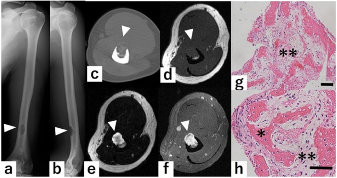
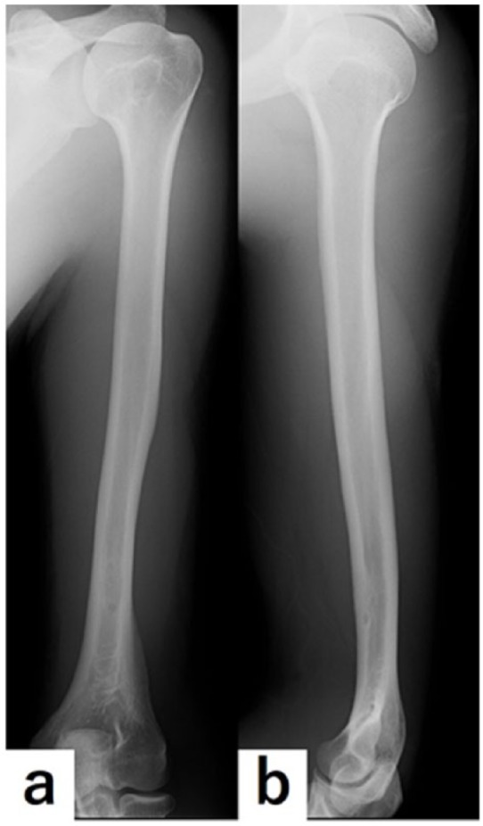
Osteofibrous dysplasia is a benign fibro-osseous lesion of bone which is most commonly occurred in cortical bone of anterior mid-shaft of the tibia of infancy and childhood. This study reported a case of osteofibrous dysplasia arising in the humerus of adult, resulting in good prognosis after a surgical treatment. A 34-year-old male had felt left upper arm pain and was suspected as having a bone tumor at the humeral shaft by X-ray pictures. The tumor was suspected as the osteofibrous dysplasia of the humerus by a core needle biopsy. Intralesional curettage, intraoperative anhydrous ethanol therapy, and artificial bone graft were performed. Surgical specimens showed fibro-osseous lesion, which strongly indicated osteofibrous dysplasia. Seven years after the surgery, he has lived without any local recurrence and complaints. It is important to recognize that osteofibrous dysplasia can arise in the humerus of an older patient for appropriate diagnosis.
Keywords: Osteofibrous dysplasia; anhydrous ethanol; humerus; ossifying fibroma.
Conflict of interest statement
Conflict of interest: The author(s) declared no potential conflicts of interest with respect to the research, authorship, and/or publication of this article.
Figures
References
-
- Fletcher CDM, Bridge JA, Hogendoorn PCW, et al. WHO classification of tumors of soft tissue and bone. 4th ed. Lyon: International Agency for Research on Cancer (IARC), 2013, pp. 343–355.
-
- Frangenheim P. Angeborene Ostitis fibrosa als Ursache einer intrauterinen Unterschenkelfraktur. Arch Klin Chir 1921; 117: 22–29 (In German).
-
- Kempson RL. Ossifying fibroma of the long bones. A light and electron microscopic study. Arch Pathol 1966; 82(3): 218–233. - PubMed
-
- Campanacci M. Osteofibrous dysplasia of long bones. A new clinical entity. Ital J Orthop Traumatol 1976; 2(2): 221–237. - PubMed
LinkOut - more resources
Full Text Sources
