

Serous cystadenoma of pancreas: A clinicopathologic experience of 23 cases from a major tertiary care center
- PMID: 30542522
- PMCID: PMC6236590
- DOI: 10.1177/2036361318809183
Serous cystadenoma of pancreas: A clinicopathologic experience of 23 cases from a major tertiary care center
Abstract
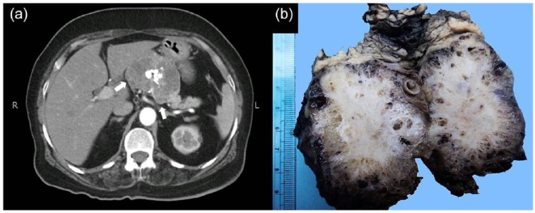
Background: Serous cystadenomas of pancreas are rare benign epithelial neoplasms, which predominantly occur in the pancreatic body and tail of elderly females. Majority of these tumors have microcystic appearance. Macrocystic and solid variants have also been described. A number of more aggressive cystic pancreatic lesions are included in the differential diagnosis. Distinction from such lesions is important for optimal management.
Objective: Our aim was to study the clinical and histological features of serous cystadenomas which would be helpful in making their correct diagnosis and understanding their behavior.
Methods: We reviewed 23 cases of serous cystadenomas diagnosed in our institution between January 2001 and June 2018.
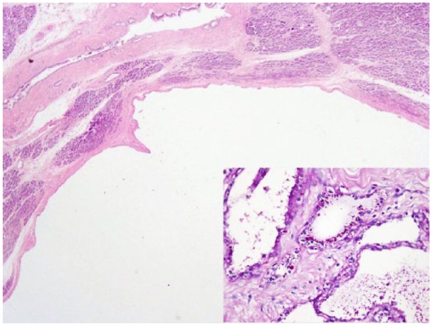
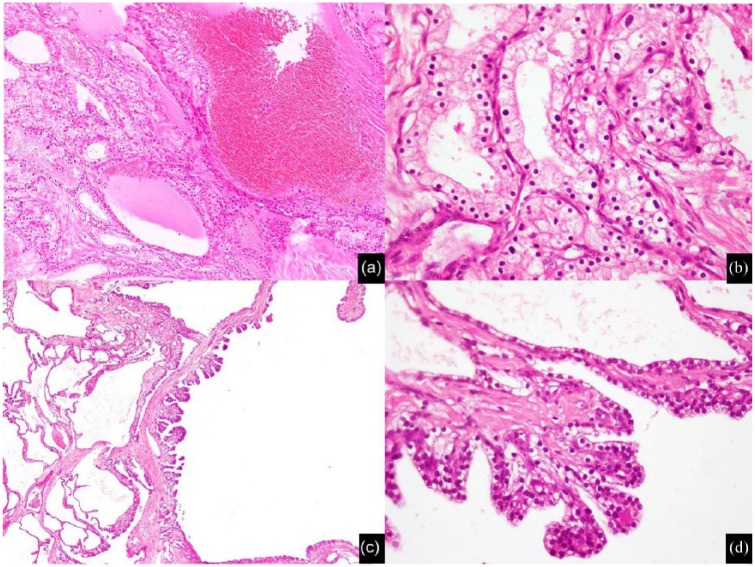
Results: Mean age at presentation was 53.43 years. Female to male ratio was 4.75:1. Over half (56.5%) of the cases were diagnosed incidentally. Abdominal pain was the most common symptom. Body and tail (either alone or in combination) were the most common locations. Tumor size ranged from 2 to 16 cm. Central scar was seen in 43.4% cases. Two cases were unilocular (macrocystic). Microscopically, all cases showed simple cuboidal to flattened epithelium with round, uniform nuclei, and glycogen-rich clear cytoplasm. Focal micropapillae formation was seen in eight cases (34.7%). Surgical resection was performed in 82.6% cases. Recurrence occurred in only one single case.
Conclusion: Pancreatic serous cystadenomas are benign neoplasms with excellent prognosis. The tumors showed typical morphological features in all cases. Surgical resection was performed in the majority of cases in our study owing to lack of optimal and complete radiological workup pre-operatively and the concern for not missing and adequately treating pancreatic mucinous cystic neoplasms.
Keywords: Serous cystadenoma; central scar; macrocystic; microcystic; pancreas.
Conflict of interest statement
Conflict of interest: The author(s) declared no potential conflicts of interest with respect to the research, authorship, and/or publication of this article.
Figures

Similar articles
-
Macrocystic pancreatic cystadenoma: The role of EUS and cyst fluid analysis in distinguishing mucinous and serous lesions.Gastrointest Endosc. 2004 Jun;59(7):823-9. doi: 10.1016/s0016-5107(04)00346-3. Gastrointest Endosc. 2004. PMID: 15173795
-
Microcystic serous cystadenoma of the pancreas with subtotal cystic degeneration: another neoplastic mimic of pancreatic pseudocyst.Am J Surg Pathol. 2012 May;36(5):726-31. doi: 10.1097/PAS.0b013e31824cf879. Am J Surg Pathol. 2012. PMID: 22498822
-
[Unilocular macrocystic serous cystadenoma of the pancreas: a morphologic variant to be considered].Ann Chir. 2003 Apr;128(3):177-9. doi: 10.1016/s0003-3944(03)00036-1. Ann Chir. 2003. PMID: 12821086 French.
-
Macrocystic serous cystadenoma of the pancreas.J Hepatobiliary Pancreat Surg. 2000;7(1):92-6. doi: 10.1007/s005340050160. J Hepatobiliary Pancreat Surg. 2000. PMID: 10982598 Review.
-
A rare case of pancreatic macrocystic serous cystadenoma in an adolescent: a case report and literature review.J Int Med Res. 2022 Oct;50(10):3000605221129102. doi: 10.1177/03000605221129102. J Int Med Res. 2022. PMID: 36259129 Free PMC article. Review.
Cited by
-
The Value of Contrast-Enhanced Ultrasound Classification in Diagnosis of Pancreatic Cystic Lesions.Biomed Res Int. 2019 Oct 13;2019:5698140. doi: 10.1155/2019/5698140. eCollection 2019. Biomed Res Int. 2019. PMID: 31737668 Free PMC article.
-
Contrast-enhanced ultrasound (CEUS) and shear wave elastography (SWE) features for characterizing serous microcystic adenomas (SMAs): In comparison to pancreatic neuroendocrine tumors (pNETs).Heliyon. 2024 Feb 1;10(3):e25185. doi: 10.1016/j.heliyon.2024.e25185. eCollection 2024 Feb 15. Heliyon. 2024. PMID: 38327470 Free PMC article.
-
Microcystic serous cystadenoma mimicking pancreatic neuroendocrine neoplasm: report of a resected case with preoperative diagnostic difficulty and review of the literature.Surg Case Rep. 2022 Sep 30;8(1):188. doi: 10.1186/s40792-022-01544-0. Surg Case Rep. 2022. PMID: 36178634 Free PMC article.
References
-
- Terris B, Fukushima N, Hruban RH. Serous neoplasms of the pancreas. In: Bosman FT, Carneiro F, Hruban RH, et al. (eds) WHO classification of tumours of the digestive system. 4th ed. Lyon: IARC Press, 2010, pp. 296–299.
-
- Hruban R, Pitman M, Klimstra D. Serous cystic neoplasms. In: Silverberg S, Sobin L. (eds) AFIP atlas of tumor pathology: tumors of the pancreas (4th series). Washington, DC: ARP Press, 2007, pp. 33–50.
-
- Colonna J, Plaza JA, Frankel WL, et al. Serous cystadenoma of the pancreas: clinical and pathological features in 33 patients. Pancreatology 2008; 8(2): 135–141. - PubMed
-
- Reid MD, Choi HJ, Memis B, et al. Serous neoplasms of the pancreas: a clinicopathologic analysis of 193 cases and review with new insights on macrocystic and solid variants and critical reappraisal of so-called “serous cystadenocarcinoma.” Am J Surg Pathol 2015; 39(12): 1597–1610. - PubMed
-
- Galanis C, Zamani A, Cameron JL, et al. Resected serous cystic neoplasms of the pancreas: a review of 158 patients with recommendations for treatment. J Gastrointest Surg 2007; 11(7): 820–826. - PubMed
LinkOut - more resources
Full Text Sources
