

Stents and Stent Mimickers in Endovascular Management of Wide-neck Intracranial Aneurysms
- PMID: 30542634
- PMCID: PMC6284878
- DOI: 10.7759/cureus.3420
Stents and Stent Mimickers in Endovascular Management of Wide-neck Intracranial Aneurysms
Abstract
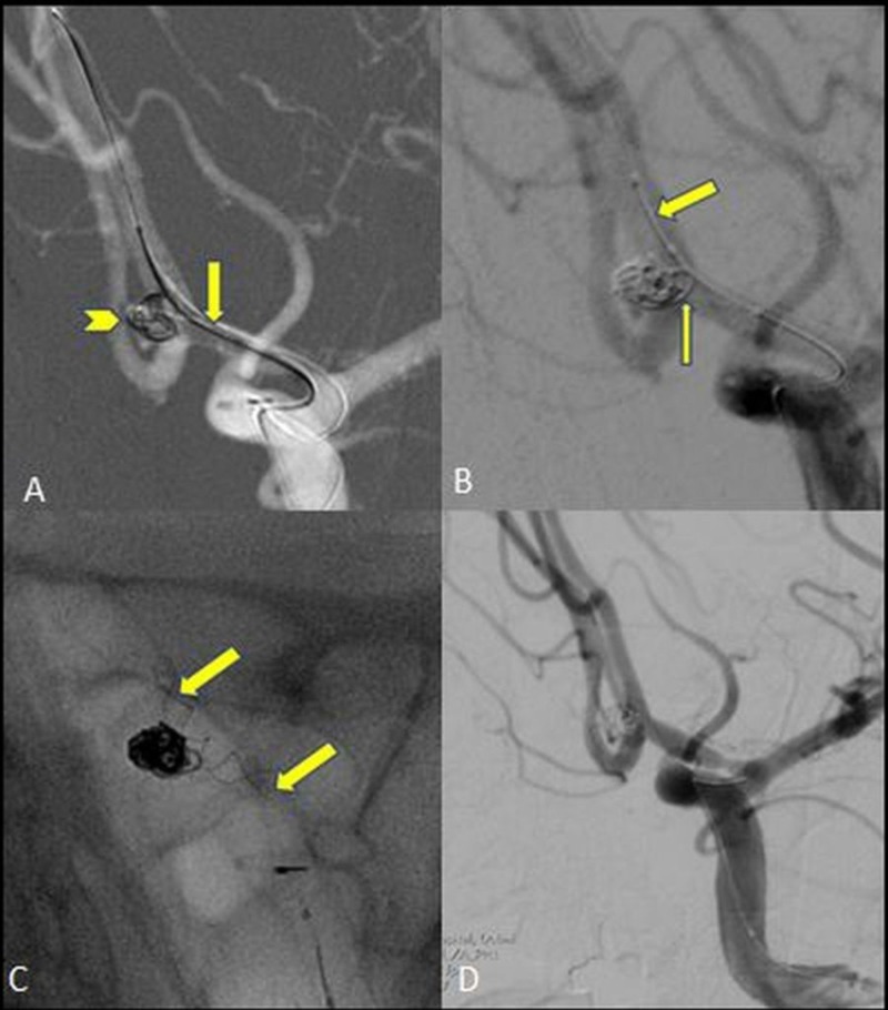
Subarachnoid hemorrhage due to a ruptured cerebral aneurysm is a disastrous event accounting for approximately 5%-15% of all stroke cases and has a high mortality rate. One of the major goals in the management of these patients is to prevent rebleeding by securing the aneurysm either surgically or by endovascular means. Endovascular treatment is considered the first line of treatment for intracranial aneurysms; however, wide-neck aneurysms (WNAs) are specifically difficult to treat by endovascular means due to the difficulty in achieving a stable coil mass inside the aneurysm sac. To overcome this problem, assisted endovascular treatment techniques and devices have evolved over the years. Amongst these, stent-assisted coiling (SAC) techniques provide a scaffold for coil embolization. The concept of the stent-assisted technique inspired creative pioneers to invent new tools like the PulseRider (Pulsar Vascular, Inc. CA, USA) and the pCONUS (Phenox GmbH, Germany), which are a great help in managing wide-neck and bifurcation aneurysms. The concept of stent within stents and its related hemodynamic effect has led to the novel development of flow diverters for reconstructing the arterial wall and correcting the hemodynamic disturbances. In this article, we review the stents and stent-like devices currently in practice for the endovascular management of wide-neck and branch intracranial aneurysms.
Keywords: coiling; endovascular; endovascular coiling; flow diversion; stent assisted; stent-assisted coiling; wide neck aneurysm.
Conflict of interest statement
The authors have declared that no competing interests exist.
Figures


Similar articles
-
Endovascular techniques for the management of wide-neck intracranial bifurcation aneurysms: A critical review of the literature.J Neuroradiol. 2016 Jun;43(3):167-75. doi: 10.1016/j.neurad.2016.02.001. Epub 2016 Mar 11. J Neuroradiol. 2016. PMID: 26976346 Review.
-
Endovascular treatment of ruptured tiny, wide-necked posterior communicating artery aneurysms using a modified stent-assisted coiling technique.J Clin Neurosci. 2013 Oct;20(10):1377-81. doi: 10.1016/j.jocn.2012.12.012. Epub 2013 Jul 23. J Clin Neurosci. 2013. PMID: 23890412
-
One-year Angiographic Results After pCONus Stent-Assisted Coiling of 40 Wide-Neck Middle Cerebral Artery Aneurysms.Neurosurgery. 2017 Jun 1;80(6):925-933. doi: 10.1093/neuros/nyw131. Neurosurgery. 2017. PMID: 28368544
-
Midterm results of T-stent-assisted coiling of wide-necked and complex intracranial bifurcation aneurysms using low-profile stents.J Neurosurg. 2017 Dec;127(6):1288-1296. doi: 10.3171/2016.9.JNS161909. Epub 2017 Jan 6. J Neurosurg. 2017. PMID: 28059656
-
Recent advances in stent-assisted coiling of cerebral aneurysms.Expert Rev Med Devices. 2020 Jun;17(6):519-532. doi: 10.1080/17434440.2020.1778463. Epub 2020 Jun 16. Expert Rev Med Devices. 2020. PMID: 32500761 Review.
Cited by
-
pCONUS 2 and pCONUS 2-HPC in the treatment of wide-necked intracranial aneurysms: Multicentre UK experience.Interv Neuroradiol. 2025 Feb;31(1):63-70. doi: 10.1177/15910199221150467. Epub 2023 Jan 8. Interv Neuroradiol. 2025. PMID: 36617807 Free PMC article.
-
Complex Wide-necked and Lobulated Aneurysm of the Middle Cerebral Artery Bifurcation : Treatment with a pCONUS2 Neck Bridging Device and p48MW Flow Modulation Device.Clin Neuroradiol. 2020 Sep;30(3):633-637. doi: 10.1007/s00062-019-00862-5. Epub 2019 Dec 5. Clin Neuroradiol. 2020. PMID: 31807809 Free PMC article. No abstract available.
-
Some technical options for successful PulseRider procedures.Surg Neurol Int. 2023 Nov 17;14:403. doi: 10.25259/SNI_656_2023. eCollection 2023. Surg Neurol Int. 2023. PMID: 38053696 Free PMC article.
-
[Clinical application of Neuroform Atlas stent-assisted coiling in the treatment of unruptured wide-neck intracranial aneurysms].Beijing Da Xue Xue Bao Yi Xue Ban. 2023 Feb 18;55(1):139-143. doi: 10.19723/j.issn.1671-167X.2023.01.021. Beijing Da Xue Xue Bao Yi Xue Ban. 2023. PMID: 36718702 Free PMC article. Chinese.
-
Comaneci-Assisted Coiling as a Treatment Option for Acutely Ruptured Wide Neck Cerebral Aneurysm: Case Series of 118 Patients.Neurosurgery. 2020 Nov 16;87(6):1148-1156. doi: 10.1093/neuros/nyaa200. Neurosurgery. 2020. PMID: 32453823 Free PMC article.
References
-
- International Subarachnoid Aneurysm Trial (ISAT) of neurosurgical clipping versus endovascular coiling in 2143 patients with ruptured intracranial aneurysms: a randomized trial. Molyneux A, Kerr R, Stratton I, et al. Lancet. 2002;360:1267–1274. - PubMed
-
- Unruptured intracranial aneurysms: natural history, clinical outcome, and risks of surgical and endovascular treatment. Wiebers DO, Whisnant JP, Huston J, et al. Lancet. 2003;362:103–110. - PubMed
-
- New detachable coils for treating cerebral aneurysms. Monsein LH. https://www.nature.com/articles/nm0296-160 Nature Medicine. 1996;2:160. - PubMed
-
- Selective endovascular treatment of 71 intracranial aneurysms with platinum coils. Casasco AE, Aymard A, Gobin YP, et al. J Neurosurg. 1993;79:3–10. - PubMed
-
- Electrothrombosis of saccular aneurysms via endovascular approach. Part 1: electrochemical basis, technique, and experimental results. Guglielmi G1, Viñuela F, Sepetka I, Macellari V. J Neurosurg. 1991;75:1–7. - PubMed
Publication types
LinkOut - more resources
Full Text Sources
Medical