. 2017 Oct;1(1):11-36.
doi: 10.15698/cst2017.10.104. Epub 2017 Oct 1.

VDAC1 at the crossroads of cell metabolism, apoptosis and cell stress

Affiliations

VDAC1 at the crossroads of cell metabolism, apoptosis and cell stress

Varda Shoshan-Barmatz et al. Cell Stress. 2017 Oct.

Abstract

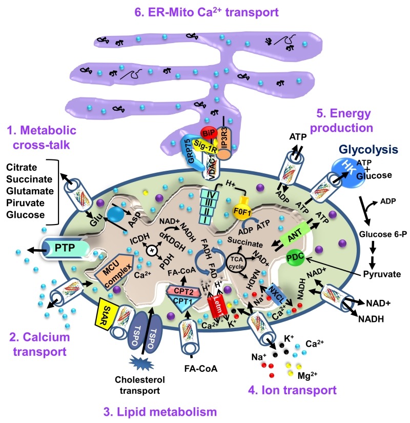
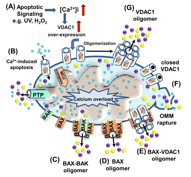
This review presents current knowledge related to VDAC1 as a multi-functional mitochondrial protein acting on both sides of the coin, regulating cell life and death, and highlighting these functions in relation to disease. It is now recognized that VDAC1 does not only play a crucial role in regulating the metabolic and energetic functions of mitochondria. The location of VDAC1 at the outer mitochondrial membrane (OMM) allows the control of metabolic cross-talk between mitochondria and the rest of the cell and also enables its interaction with proteins involved in metabolic and survival pathways. Along with regulating cellular energy production and metabolism, VDAC1 is also involved in the process of mitochondria-mediated apoptosis by mediating the release of apoptotic proteins and interacting with anti-apoptotic proteins. VDAC1 functions in the release of apoptotic proteins located in the mitochondrion inter-membranal space via oligomerization to form a large channel that allows passage of cytochrome c and AIF and their release to the cytosol, subsequently apoptotic cell death. VDAC1 also regulates apoptosis via interactions with apoptosis regulatory proteins, such as hexokinase (HK), Bcl2 and Bcl-xL, some of which are also highly expressed in many cancers. This review also provide insight into VDAC1 function in Ca2+ homeostasis, oxidative stress, and presents VDAC1 as a hub protein interacting with over 100 proteins. Such interactions enable VDAC1 to mediate and regulate the integration of mitochondrial functions with cellular activities. VDAC1 can thus be considered as standing at the crossroads between mitochondrial metabolite transport and apoptosis and hence represents an emerging cancer drug target.

Keywords: Apoptosis; Cancer; Metabolism; Mitochondria; VDAC1.


Conflict of interest statement

Conflict of interest: The authors declare no conflict of interest.

Figures

Figure 1
Figure 1. FIGURE 1: Three-dimensional structure of VDAC1.
VDAC1 monomer and dimer structures. (A) Side-view of the crystal structure of VDAC1 (PDB code: 3EMN). The β-barrel is formed by 19 β strands and the N-terminal domain (colored red) is folded into the pore interior. (B) A proposed model for the conformation of VDAC1 with its N-terminal on the outside of the VDAC1 pore. (C) Top-view of VDAC1 dimer with the N-terminal helix nested inside the VDAC1 pore in one monomer and outside of the pore in the other. (D) Side-view of proposed dimer of VDAC1. Figures were prepared using PyMOL software.
Figure 2
Figure 2. FIGURE 2: VDAC1 as a multi-functional channel involved in metabolite, cholesterol and Ca2+ transport, energy production and in ER-mitochondria structural and functional association.
The functions of VDAC1 in cell life include control of the metabolic cross-talk between the mitochondria and the rest of the cell energy production, regulation of glycolysis via binding of HK, Ca2+ signaling and cholesterol transport. The various functions of VDAC1 in the cell and mitochondria functions are presented. These include: 1. Control of the metabolic cross-talk between mitochondria and the rest of the cell; 2. Transport of Ca2+ to and from the IMS and acting in Ca2+ signaling; 3. Lipid metabolism; 4. Transport of ions, such as Mg2+, Zn+, Na+ and K+; 5. Mediating cellular energy production by transporting ATP/ADP and NAD+/NADH and acyl-CoA (FA-CoA) from the cytosol to and from the IMS, and regulating glycolysis via association with HK; 6. Structurally and functionally contributing to ER-mitochondria contacts, mediating Ca2+ transport from the ER to mitochondria. Ca2+ influx and efflux systems in the IMM are shown. The mitochondrial Ca2‏+ uniporter (MCU), in association with a calcium-sensing accessory subunit (MCU1), mediates Ca2+ transport from the IMS into the matrix. The ryanodine receptor (RyR) in the IMM mediates Ca2+ influx. NCLX, a Na+/Ca2+ exchanger, mediates Ca2+ efflux from the matrix to the IMS. High levels of matrix Ca2‏+ trigger the opening of the PTP, a fast Ca2+ release channel. Molecular fluxes are indicated by arrows. The function of Ca2+ in regulating energy production is mediated via activation of the TCA cycle enzymes pyruvate dehydrogenase (PDH), isocitrate dehydrogenase (ICDH) and α-ketoglutarate dehydrogenase (α-KGDH), leading to enhanced activity of the TCA cycle. The electron transport chain (ETC) and the ATP synthase (FoF1) are also presented. VDAC1 mediates the transfer of acyl-CoAs across the OMM to the IMS, where they are converted into acylcarnitine by CPT1a for further processing by β-oxidation. VDAC1 is involved in cholesterol transport by being a constituent of a multi-protein complex, the transduceosome, containing StAR/TSPO/VDAC1.The ER-mitochondria association is presented with key proteins indicated. These include the inositol 3 phosphate receptor type 3 (IP3R3), the sigma 1 receptor (Sig1R) (a reticular chaperone), binding immunoglobulin protein (BiP), the ER HSP70 chaperone, and glucose-regulated protein 75 (GRP75). IP3 activates IP3R in the ER to release Ca2+ that is directly transferred to the mitochondria via VDAC1.
Figure 3
Figure 3. FIGURE 3: VDAC1-tubulin interaction: a metabolic switch to modulate mitochondrial metabolism in cancer cells.
In cancer cells, high levels of free tubulin close VDAC1, decreasing the flux of metabolites, ATP and ADP through the OMM. VDAC1 closing leads to low generation of mitochondrial ATP and subsequently, to a low cytosolic ATP/ADP ratio that favors glycolysis in the Warburg phenotype. Erastin, a VDAC-tubulin antagonist, opens VDAC1 by blocking the inhibitory effect of free tubulin. VDAC1 opening leads to increased mitochondrial metabolism and to a high cytosolic ATP/ADP ratio that inhibits glycolysis and reverts the Warburg phenotype. αβ indicates tubulin heterodimers.
Figure 4
Figure 4. FIGURE 4: VDAC1-depletion and metabolic reprogramming leading to alterations in key transcription factor levels and biological processes: a reversal of oncogenic properties and cell differentiation.
(A) A schematic presentation of mitochondria in a cancer cell before treatment with hVDAC1 siRNA. Here, cancer cells maintain homeostatic energy and metabolic states, with HK bound to VDAC1 accelerating glycolysis and mitochondrial function to allow sufficient ATP and metabolite precursor levels to support cell growth and survival. (B) VDAC1 depletion leads to dramatic decreases in energy and metabolite generation. This leads to changes in master metabolism regulator (p53, HIF1-α, c-Myc and NF-kb, P65) expression levels, which alters the expression of transcription factors associated with stemness, EMT, cell proliferation, invasion, TAMs and angiogenesis, while leading to differentiation into astrocyte- or neuron-like cells.
Figure 5
Figure 5. FIGURE 5: VDAC1 function in cell death, with apoptosis inducers enhancing VDAC1 expression levels and oligomerization.
A schematic representation of VDAC1 function in cell death - Different models for the release of apoptogenic proteins, such as Cyto c (purple) and AIF (yellow), are shown. (A) Proposed model suggesting that apoptotic stimuli or conditions cause enhanced VDAC1 expression via increases in [Ca2+]i levels or transcription factors, leading to activation of the VDAC1 promoter. The increase in VDAC1 expression shifts the equilibrium towards the VDAC1 oligomeric state, forming a hydrophilic protein-conducting channel capable of mediating the release of apoptogenic proteins (e.g., Cyto c and AIF) from the mitochondrial IMS to the cytosol. (B) Mitochondrial Ca2+ overload induces apoptosis. Ca2+ transport across the OMM, as mediated by VDAC1, and then across the IMM, as mediated by the MCU, leads to Ca2+ overload in the matrix. This, in turn, causes dissipation of the membrane potential, mitochondrial swelling, PTP opening, Cyto c/AIF release and the triggering of apoptotic cell death. (C) Bax/Bak oligomerization and activation, forming a route for Cyto c/AIF release. (D) Bax activation leads to its association with the OMM, followed by its oligomerization as a large oligomer/complex, forming a Cyto c/AIF-conducting channel. (E) The interaction of the pro-apoptotic protein Bax with VDAC1 forms hetro-oligomers that mediate Cyto c/AIF release. (F) Prolonged VDAC1 closure leads to mitochondrial matrix swelling and OMM rupture, resulting in the appearance of a non-specific release pathway for apoptogenic proteins.

Similar articles

Cited by

References

    1. Shoshan-Barmatz V, De Pinto V, Zweckstetter M, Raviv Z, Keinan N, Arbel N. VDAC, a multi-functional mitochondrial protein regulating cell life and death. Mol Aspects Med. 2010;31(3):227–285. doi: 10.1016/j.mam.2010.03.002. - DOI - PubMed
    1. Messina A, Reina S, Guarino F, De Pinto V. VDAC isoforms in mammals. Biochim Biophys Acta. 2012;1818(6):1466–1476. doi: 10.1016/j.bbamem.2011.10.005. - DOI - PubMed
    1. De Pinto V, Guarino F, Guarnera A, Messina A, Reina S, Tomasello FM, Palermo V, Mazzoni C. Characterization of human VDAC isoforms: a peculiar function for VDAC3? Biochim Biophys Acta. 2010;1797(6-7):1268–1275. doi: 10.1016/j.bbabio.2010.01.031. - DOI - PubMed
    1. De Pinto V, Reina S, Gupta A, Messina A, Mahalakshmi R. Role of cysteines in mammalian VDAC isoforms' function. Biochim Biophys Acta. 2016;1857(8):1219–1227. doi: 10.1016/j.bbabio.2016.02.020. - DOI - PMC - PubMed
    1. Cheng EH, Sheiko TV, Fisher JK, Craigen WJ, Korsmeyer SJ. VDAC2 inhibits BAK activation and mitochondrial apoptosis. 2003;(5632):513–517. doi: 10.1126/science.1083995. - DOI - PubMed

LinkOut - more resources