Structure, asymmetry, and connectivity of the human temporo-parietal aslant and vertical occipital fasciculi
- PMID: 30542766
- PMCID: PMC7026858
- DOI: 10.1007/s00429-018-1812-0
Structure, asymmetry, and connectivity of the human temporo-parietal aslant and vertical occipital fasciculi
Abstract
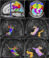
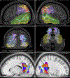
We previously proposed a bipartite 'dorsal-ventral' model of human arcuate fasciculus (AF) morphology. This model does not, however, account for the 'vertical,' temporo-parietal subdivision of the AF described in earlier dissection and tractographic studies. In an effort to address the absence of the vertical AF (VAF) within 'dorsal-ventral' nomenclature, we conducted a dedicated tractographic and white-matter dissection study of this tract and another short, vertical, posterior-hemispheric fascicle: the vertical occipital fasciculus (VOF). We conducted atlas-based, non-tensor, deterministic tractography in 30 single subjects from the Human Connectome Project database and verified our results using an average diffusion atlas compiled from 842 separate normal subjects. We also performed white-matter dissection in four post-mortem specimens. Our tractography results demonstrate that the VAF is, in fact, a bipartite system connecting the ventral parietal and temporal regions, with variable connective, and no volumetric lateralization. The VOF is a non-lateralized, non-segmented system connecting lateral occipital areas with basal-temporal regions. Importantly, the VOF was spatially dissociated from the VAF. As the VAF demonstrates no overall connective or volumetric lateralization, we postulate its distinction from the AF system and propose its re-naming to the 'temporo-parietal aslant tract,' (TPAT), with unique dorsal and ventral subdivisions. Our tractography results were supported by diffusion atlas and white-matter dissection findings.
Conflict of interest statement
Disclosure
Authors report no conflicts of interest.
Figures


Similar articles
-
Identification of a distinct association fiber tract "IPS-FG" to connect the intraparietal sulcus areas and fusiform gyrus by white matter dissection and tractography.Sci Rep. 2020 Sep 23;10(1):15475. doi: 10.1038/s41598-020-72471-z. Sci Rep. 2020. PMID: 32968114 Free PMC article.
-
Mapping temporo-parietal and temporo-occipital cortico-cortical connections of the human middle longitudinal fascicle in subject-specific, probabilistic, and stereotaxic Talairach spaces.Brain Imaging Behav. 2017 Oct;11(5):1258-1277. doi: 10.1007/s11682-016-9589-3. Brain Imaging Behav. 2017. PMID: 27714552 Free PMC article.
-
A Connectomic Atlas of the Human Cerebrum-Chapter 16: Tractographic Description of the Vertical Occipital Fasciculus.Oper Neurosurg. 2018 Dec 1;15(suppl_1):S456-S461. doi: 10.1093/ons/opy270. Oper Neurosurg. 2018. PMID: 30260427 Free PMC article.
-
The vertical superior longitudinal fascicle and the vertical occipital fascicle.J Neurosurg Sci. 2021 Dec;65(6):581-589. doi: 10.23736/S0390-5616.21.05368-6. J Neurosurg Sci. 2021. PMID: 35128919 Review.
-
Anatomical variability of the arcuate fasciculus: a systematical review.Surg Radiol Anat. 2019 Aug;41(8):889-900. doi: 10.1007/s00276-019-02244-5. Epub 2019 Apr 26. Surg Radiol Anat. 2019. PMID: 31028450
Cited by
-
Association fiber tracts related to Broca's area: A comparative study based on diffusion spectrum imaging and fiber dissection.Front Neurosci. 2022 Nov 7;16:978912. doi: 10.3389/fnins.2022.978912. eCollection 2022. Front Neurosci. 2022. PMID: 36419463 Free PMC article.
-
Intraoperative augmented reality fiber tractography complements cortical and subcortical mapping.World Neurosurg X. 2023 Jun 23;20:100226. doi: 10.1016/j.wnsx.2023.100226. eCollection 2023 Oct. World Neurosurg X. 2023. PMID: 37456694 Free PMC article.
-
Neural Connectivity: How to Reinforce the Bidirectional Synapse Between Basic Neuroscience and Routine Neurosurgical Practice?Front Neurol. 2021 Jul 1;12:705135. doi: 10.3389/fneur.2021.705135. eCollection 2021. Front Neurol. 2021. PMID: 34354668 Free PMC article. No abstract available.
-
Occipital Intralobar fasciculi: a description, through tractography, of three forgotten tracts.Commun Biol. 2021 Mar 30;4(1):433. doi: 10.1038/s42003-021-01935-3. Commun Biol. 2021. PMID: 33785859 Free PMC article.
-
Inter-individual Differences in Occipital Alpha Oscillations Correlate with White Matter Tissue Properties of the Optic Radiation.eNeuro. 2020 Apr 24;7(2):ENEURO.0224-19.2020. doi: 10.1523/ENEURO.0224-19.2020. Print 2020 Mar/Apr. eNeuro. 2020. PMID: 32156741 Free PMC article.
References
MeSH terms
Grants and funding
LinkOut - more resources
Full Text Sources
Research Materials

