. 2019 Feb;24(2):121-132.
doi: 10.1177/2472555218813332. Epub 2018 Dec 13.

Positioning High-Throughput CETSA in Early Drug Discovery through Screening against B-Raf and PARP1

Affiliations

Positioning High-Throughput CETSA in Early Drug Discovery through Screening against B-Raf and PARP1

Joseph Shaw et al. SLAS Discov. 2019 Feb.

Abstract

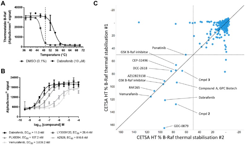
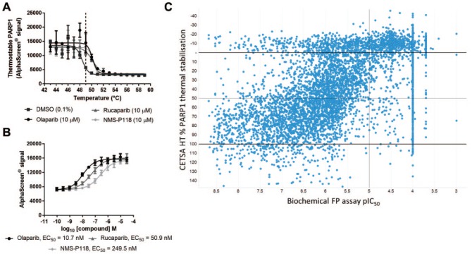
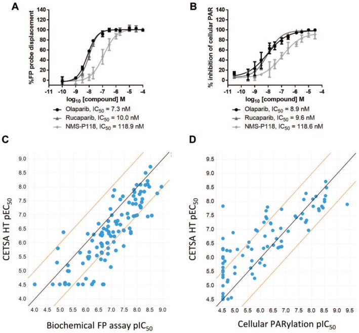
Methods to measure cellular target engagement are increasingly being used in early drug discovery. The Cellular Thermal Shift Assay (CETSA) is one such method. CETSA can investigate target engagement by measuring changes in protein thermal stability upon compound binding within the intracellular environment. It can be performed in high-throughput, microplate-based formats to enable broader application to early drug discovery campaigns, though high-throughput forms of CETSA have only been reported for a limited number of targets. CETSA offers the advantage of investigating the target of interest in its physiological environment and native state, but it is not clear yet how well this technology correlates to more established and conventional cellular and biochemical approaches widely used in drug discovery. We report two novel high-throughput CETSA (CETSA HT) assays for B-Raf and PARP1, demonstrating the application of this technology to additional targets. By performing comparative analyses with other assays, we show that CETSA HT correlates well with other screening technologies and can be applied throughout various stages of hit identification and lead optimization. Our results support the use of CETSA HT as a broadly applicable and valuable methodology to help drive drug discovery campaigns to molecules that engage the intended target in cells.

Keywords: B-Raf; CETSA; PARP1; target engagement.


Conflict of interest statement

Declaration of Conflicting Interests: The authors declared the following potential conflicts of interest with respect to the research, authorship, and/or publication of this article: J.S., I.D., P.H., L.L., J.P.O., V.T., A.J.N., M.B., M.J.M., and D.G. are employees of AstraZeneca and potentially own stock and/or stock options in the company. N.D., D.M.M., and M.D. are employees of Pelago Bioscience AB and own stock in the company.

Figures

Figure 1.
Figure 1.
A CETSA HT assay to screen for intracellular B-Raf target engagement. (A) A375 cells were treated for 1 h with DMSO control or dabrafenib (10 µM) before applying indicated heatshock, lysis, and quantification of thermostable B-Raf by AlphaScreen. Treatment of live cells with dabrafenib caused a thermal stabilization of B-Raf. Data are the mean ± span of n = 2. (B) ITDRFCETSA experiments to rank intracellular B-Raf target engagement. A375 cells were treated with a concentration response of B-Raf inhibitors for 2 h prior to a 49 °C heatshock, lysis, and quantification of thermostable B-Raf by AlphaScreen. Data are the mean ± standard deviation of n = 4. (C) CETSA HT screening of a library of kinase inhibitors to identify in-cell B-Raf binders. Compounds were screened at a test concentration of 30 µM for thermal stabilization of B-Raf relative to the dabrafenib control following 2 h incubation with A375 cells across two technical repeats.
Figure 2.
Figure 2.
A CETSA HT assay to screen for intracellular PARP1 target engagement. (A) MDA-MB-436 cells were treated for 1 h with DMSO, olaparib (10 µM), rucaparib (10 µM), or NMS-P118 (10 µM) before applying indicated heatshock, lysis, and quantification of thermostable PARP1 by AlphaScreen. Treatment of live cells with PARP inhibitors led to a thermal stabilization of PARP1. Data are the mean ± span of n = 2. (B) ITDRFCETSA experiments to rank intracellular PARP1 target engagement. MDA-MB-436 cells were treated with a concentration response of PARP inhibitors for 1 h prior to a 49 °C heatshock, lysis, and quantification of thermostable PARP1 by AlphaScreen. Thermal stabilization as a measure of target engagement allowed compounds to be ranked by apparent potency. Data are the mean ± standard deviation of n = 10. (C) Single-concentration CETSA HT screening of a library of PARP1 binders. The affinity (pIC50) of 6288 compounds to PARP1 protein was determined by concentration–response experiments using a biochemical FP assay (x axis). The same compounds were screened for CETSA HT thermal stabilization at 10 µM, plotted as percent PARP1 thermal stabilization relative to 100% olaparib stabilized (y axis).
Figure 3.
Figure 3.
Comparison of PARP inhibitor potency in CETSA HT with alternative assay formats. (A) A biochemical FP assay was employed to measure binding to purified PARP1 protein via competition of an FP probe. Data for PARP inhibitors, comparable to CETSA data in Figure 2B, are shown. Data are the mean ± standard deviation of ⩾4 technical replicates. (B) A cellular PARylation assay was employed to measure cellular PARP function. A549 cells were treated with PARP inhibitors for 1 h before addition of a DNA-damaging agent and imaging and quantifying PARylation. Data are the mean ± standard deviation of four technical replicates. (C) pEC50 determined by ITDRFCETSA plotted against pIC50 determined in the biochemical FP assay for 112 PARP inhibitors. The solid line shows a 1:1 correlation, and dashed lines represent a 1 log10 shift in potency. (D) pEC50 determined by ITDRFCETSA plotted against pIC50 determined in the cellular PARylation assay for 99 of the same PARP inhibitors. The solid line shows a 1:1 correlation, and dashed lines represent a 1 log10 shift in potency.

References

    1. Simon G. M., Niphakis M. J., Cravatt B. F. Determining Target Engagement in Living Systems. Nat. Chem. Biol. 2013, 9, 200–205. - PMC - PubMed
    1. Morgan P., Brown D. G., Lennard S., et al. Impact of a Five-Dimensional Framework on R&D Productivity at AstraZeneca. Nat. Rev. Drug Discov. 2018, 17, 167–181. - PubMed
    1. Smyth L. A., Collins I. Measuring and Interpreting the Selectivity of Protein Kinase Inhibitors. J. Chem. Biol. 2009, 2, 131–151. - PMC - PubMed
    1. Martinez Molina D., Jafari R., Ignatushchenko M., et al. Monitoring Drug Target Engagement in Cells and Tissues Using the Cellular Thermal Shift Assay. Science 2013, 341, 84–87. - PubMed
    1. Almqvist H., Axelsson H., Jafari R., et al. CETSA Screening Identifies Known and Novel Thymidylate Synthase Inhibitors and Slow Intracellular Activation of 5-Fluorouracil. Nat. Commun. 2016, 7, 11040. - PMC - PubMed

MeSH terms

Substances