

Release of fascial compartment boundaries reduces muscle force output
- PMID: 30543496
- PMCID: PMC6459388
- DOI: 10.1152/japplphysiol.00330.2018
Release of fascial compartment boundaries reduces muscle force output
Abstract
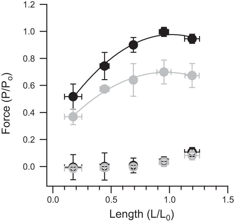
Most limb muscles operate within a compartment defined by fascial layers that enclose a muscle or groups of muscles within a defined space. These compartments are important clinically, because fluid accumulation can cause ischemia and tissue necrosis if untreated. Little is known, however, about how fascial enclosures influence healthy muscle function. One previous study showed that removing a fascial covering reduced the force output of a muscle under maximal stimulation. We hypothesized that such reduction in force output was due to a change in the muscle length following fasciotomy and that a reduced force output could be explained by the length-tension relationship of muscle. Thus we predicted that the maximum force across a range of lengths would be unchanged following fasciotomy. We measured maximal tetanic force output in a wing muscle in wild turkeys both before and after removal of fascia that enclosed the muscle in a compartment. Our hypothesis was not supported. The length-tension curve of this muscle showed that removal of fascia reduced maximum force output to 72 ± 10% of the prefascial release condition. Thus a reduction in muscle force following fasciotomy was not explained by a change in muscle length. The mechanism underlying reduction in force is unclear, but it suggests that the assumption underlying most isolated muscle experiments, i.e., removal of a muscle from its situation in vivo does not influence its maximal mechanical output, may need reexamining. NEW & NOTEWORTHY Most limb muscles are enclosed within compartments bound by robust fascial sheets. The mechanical significance of the close packing of muscle and fascia is largely unexplored. We used an animal model to show that removal of a fascial covering reduces the maximal force developed during contraction. These results raise questions about the use of isolated muscles to estimate muscle performance and suggest that a muscle's mechanical surrounding influences performance by mechanisms that are not understood.
Keywords: compartment; fasciotomy; intramuscular pressure; muscle; tendon.
Conflict of interest statement
No conflicts of interest, financial or otherwise, are declared by the authors.
Figures


Similar articles
-
Myofascial force transmission causes interaction between adjacent muscles and connective tissue: effects of blunt dissection and compartmental fasciotomy on length force characteristics of rat extensor digitorum longus muscle.Arch Physiol Biochem. 2001 Apr;109(2):97-109. doi: 10.1076/apab.109.2.97.4269. Arch Physiol Biochem. 2001. PMID: 11780782
-
Compartmental fasciotomy and isolating a muscle from neighboring muscles interfere with myofascial force transmission within the rat anterior crural compartment.J Morphol. 2003 Jun;256(3):306-21. doi: 10.1002/jmor.10097. J Morphol. 2003. PMID: 12655613
-
Transmission of muscle force to fascia during exercise.J Bodyw Mov Ther. 2015 Jan;19(1):119-23. doi: 10.1016/j.jbmt.2014.08.010. Epub 2014 Sep 3. J Bodyw Mov Ther. 2015. PMID: 25603751
-
Fascial hierarchies and the relevance of crossed-helical arrangements of collagen to changes in the shape of muscles.J Bodyw Mov Ther. 2016 Apr;20(2):377-87. doi: 10.1016/j.jbmt.2015.09.004. Epub 2015 Oct 23. J Bodyw Mov Ther. 2016. PMID: 27210857 Review.
-
Force transmission between synergistic skeletal muscles through connective tissue linkages.J Biomed Biotechnol. 2010;2010:575672. doi: 10.1155/2010/575672. Epub 2010 Apr 12. J Biomed Biotechnol. 2010. PMID: 20396618 Free PMC article. Review.
Cited by
-
Muscle Morphology Does Not Solely Determine Knee Flexion Weakness After Anterior Cruciate Ligament Reconstruction with a Semitendinosus Tendon Graft: A Combined Experimental and Computational Modeling Study.Ann Biomed Eng. 2024 May;52(5):1313-1325. doi: 10.1007/s10439-024-03455-7. Epub 2024 Feb 29. Ann Biomed Eng. 2024. PMID: 38421479 Free PMC article.
-
Effect of muscle stimulation intensity on the heterogeneous function of regions within an architecturally complex muscle.J Appl Physiol (1985). 2021 Apr 1;130(4):941-951. doi: 10.1152/japplphysiol.00514.2020. Epub 2021 Jan 7. J Appl Physiol (1985). 2021. PMID: 33411643 Free PMC article.
-
Force enhancement in the human vastus lateralis is muscle-length-dependent following stretch but not during stretch.Eur J Appl Physiol. 2020 Dec;120(12):2597-2610. doi: 10.1007/s00421-020-04488-1. Epub 2020 Sep 5. Eur J Appl Physiol. 2020. PMID: 32892321 Free PMC article.
-
Hindlimb muscle spindles inform preparatory forelimb coordination prior to landing in toads.J Exp Biol. 2023 Jan 15;226(2):jeb244629. doi: 10.1242/jeb.244629. Epub 2023 Jan 19. J Exp Biol. 2023. PMID: 36576050 Free PMC article.
-
Internal fluid pressure influences muscle contractile force.Proc Natl Acad Sci U S A. 2020 Jan 21;117(3):1772-1778. doi: 10.1073/pnas.1914433117. Epub 2019 Dec 26. Proc Natl Acad Sci U S A. 2020. PMID: 31879350 Free PMC article.
References
-
- Askew GN, Marsh RL. The effects of length trajectory on the mechanical power output of mouse skeletal muscles. J Exp Biol 200: 3119–3131, 1997. - PubMed
-
- Brown DE. The effect of rapid changes in hydrostatic pressure upon the contraction of skeletal muscle. J Cell Comp Physiol 4: 257–281, 1934. doi:10.1002/jcp.1030040208. - DOI
-
- Cattell M, Edwards DJ. The energy changes of skeletal muscle accompanying contraction under high pressure. Am J Physiol 86: 371–382, 1928. doi:10.1152/ajplegacy.1928.86.2.371. - DOI
Publication types
MeSH terms
Grants and funding
LinkOut - more resources
Full Text Sources