. 2018 Dec 13;13(12):e0198271.
doi: 10.1371/journal.pone.0198271. eCollection 2018.

The neuroanatomy of the siboglinid Riftia pachyptila highlights sedentarian annelid nervous system evolution

Affiliations

The neuroanatomy of the siboglinid Riftia pachyptila highlights sedentarian annelid nervous system evolution

Nadezhda N Rimskaya-Korsakova et al. PLoS One. .

Abstract

Tracing the evolution of the siboglinid group, peculiar group of marine gutless annelids, requires the detailed study of the fragmentarily explored central nervous system of vestimentiferans and other siboglinids. 3D reconstructions of the neuroanatomy of Riftia revealed that the "brain" of adult vestimentiferans is a fusion product of the supraesophageal and subesophageal ganglia. The supraesophageal ganglion-like area contains the following neural structures that are homologous to the annelid elements: the peripheral perikarya of the brain lobes, two main transverse commissures, mushroom-like structures, commissural cell cluster, and the circumesophageal connectives with two roots which give rise to the palp neurites. Three pairs of giant perikarya are located in the supraesophageal ganglion, giving rise to the paired giant axons. The circumesophageal connectives run to the VNC. The subesophageal ganglion-like area contains a tripartite ventral aggregation of perikarya (= the postoral ganglion of the VNC) interconnected by the subenteral commissure. The paired VNC is intraepidermal, not ganglionated over most of its length, associated with the ciliary field, and comprises the giant axons. The pairs of VNC and the giant axons fuse posteriorly. Within siboglinids, the vestimentiferans are distinguished by a large and considerably differentiated brain. This reflects the derived development of the tentacle crown. The tentacles of vestimentiferans are homologous to the annelid palps based on their innervation from the dorsal and ventral roots of the circumesophageal connectives. Neuroanatomy of the vestimentiferan brains is close to the brains of Cirratuliiformia and Spionida/Sabellida, which have several transverse commissures, specific position of the giant somata (if any), and palp nerve roots (if any). The palps and palp neurite roots originally developed in all main annelid clades (basally branching, errantian and sedentarian annelids), show the greatest diversity in their number in sedentarian species. Over the course of evolution of Sedentaria, the number of palps and their nerve roots either dramatically increased (as in vestimentiferan siboglinids) or were lost.


Conflict of interest statement

The authors have declared that no competing interests exist.

Figures

Fig 1
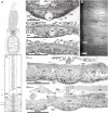
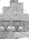
Fig 1. The anterior ventral nerve cord within the vestimental collar of Riftia pachyptila.
In the anteriormost part of the vestimentum, the ventral brain connects to the ventral nerve cord (A, B). The longitudinal nerve tracts (LNT) are continuations of the nerve fibres running from the ventral nerve cord to the posterior and anterior parts of the brain (D, C). The nerve cord lies completely inside the epidermis (E). A—scheme of the central nervous system: main elements in grey, giant axons in light grey. Anterior ends at top. Frame indicates the area corresponding to transverse (B-D) and parasagittal (E) sections. Stars show the levels of the corresponding transverse sections. Parasagittal section (E) made through one of the paired strands of the VNC. Dotted lines show region borders. B—posteriormost brain; elements of ventral nerve cord projecting into brain, juvenile specimen. C–ventral nerve cord (VNC) directly posterior to brain, adult specimen. D–longitudinal nerves at transition of ventral nerve cord and brain. E–intraepidermal ventral nerve cord (VNC). B–brain, CNC–commissural neurite bundles of VNC, cpnc–central perikarya of VNC, CU–cuticle, ECM–extracellular matrix, EP–epidermis, EXP–epidermal cell processes, GA–giant axons, EC–enteral coelom, LN–circular neurite bundles, lpnc–lateral perikarya of VNC, lvtp–ventrolateral perikarya of tripartite ventral aggregation, NE–neuropile of lateral brain lobes, nep–peripheral perikarya of lateral brain lobes, OB–obturaculum, OBC–obturacular coelom, OP–opisthosome, pmp–posterior median perikarya aggregation, PNC–paired strands of VNC surrounding ventral ciliary field, vvtp–ventral perikarya of tripartite ventral aggregation, TR–trunk, VNC–ventral nerve cord, VT–vestimentum, VWF–collar of vestimental wings, VWN–neurite bundle of VW.
Fig 2
Fig 2. The ventral nerve cord in the vestimentum of Riftia.
In the vestimentum, the paired strands of the ventral nerve cord (VNC) split and surround the ciliary field (CF, A-C). The pair of giant axons lies in each strand of the VNC (C, D). Numerous lateral neurite bundles arise from the nerve strands (E). The strands and giant axons unite at the boarder between the vestimentum and trunk (A). A–scheme of central nervous system: main elements in grey, giant axons in light grey. Anterior ends at top. Frame indicates area corresponding to histological cross sections (B-E). Stars show the levels of the corresponding sections. B–ventral nerve cord directly anteriorly to ventral ciliary field. C–ventral ciliary field (CF) surrounded by paired strands of ventral nerve cord (PNC); line shows border of ciliary field, brackets show strands of ventral nerve cord. D–close-up of left strand of ventral nerve cord, commissural neurite bundles connecting the paired strands are visible (CNC). E–lateral circular neurite bundles (LN) in epidermis. F–one of the lateral neurite bundles running in the body wall epidermis. B–brain, BV–blood vessels, CNC–commissural neurite bundles of VNC, CM–circular musculature, CF–ventral ciliary field, CIL–cilia, cpnc–central perikarya of VNC, CU–cuticle, ECM–extracellular matrix, ega–cells coating GA, EP–epidermis, GA–giant axons, LN–circular neurite bundles, lpnc–lateral perikarya of VNC, NNC–neuropile of VNC, OB–obturaculum, OP–opisthosome, PNC–paired strands of VNC surrounding CF, TR–trunk, VNC–ventral nerve cord, VT–vestimentum.
Fig 3
Fig 3. The ventral nerve cord in the trunk and opisthosome of Riftia.
The trunk contains a single ventral nerve cord (VNC) (A) whose diameter gradually decreases towards the end of the trunk (B-D). A single giant axon extends to the border of the trunk and opisthosome (A-D). Numerous lateral neurite bundles (LN) arises from the VNC in the trunk, whereas in each segment of the opisthosome, only one pair of LN arises from the VNC (E-G). A–scheme of central nervous system: main elements in grey, giant axons in light grey. Anterior ends at top. Frame indicates area corresponding to histological sections (B-D, F, G) and light miscroscopical image (E). Stars show the levels of the corresponding sections. B–ventral nerve cord (VNC) structure in anterior trunk. C, D–VNC structure in mid- and posterior trunk, respectively; note reduction of giant axon diameter. E–lateral neurite bundles branching and anastomosing in trunk epidermis, anterior ends at top. F-G–VNC in mid- and posterior opisthosome. Arrows in (F): cuticular folds between cell borders. ECM–extracellular matrix, BV–blood vessels, CM–circular muscles, D–dissepiments, ega–cells coating GA, EP–epidermis, EXP–epidermal cell processes, GA–giant axons, FM–featherlike longitudinal muscles, LG–longitudinal lateral grooves, LM–longitudinal muscles, LN–circular neurite bundles, lpnc–lateral perikarya of VNC, ME–mesenterium, NNC–neuropile of VNC, OP–opisthosome, PA–cuticular plaque papillae, pl–large perikarya, ps–small perikarya, TR–trunk, VNC–ventral nerve cord.
Fig 4
Fig 4. Brain of juvenile Riftia with a gut rudiment.
Brain of R. pachyptila consists of dorsal and ventral parts divided by the enteral coelom (EC) containing the gut (G) (A, B). The dorsal part is associated with the supraesophageal ganglion, the ventral part with the subesophageal ganglion. A–idealised scheme of sagittal section of vestimentiferan brain, which consists of supraesophageal and subesophageal ganglia divided by the enteral coelom (EC). Anterior ends to left. B–one of the parasagittal sections of 8-mm-long juvenile; gut rudiment passes through brain. Anterior ends to left. amp–anterior median aggregation of perikarya, CUP–cuticle schield, DC–dorsal commissure, DLN–dorsal longitudinal bundles, ET–excretory tree, G–gut lumen, EC–enteral coelom, H–heart, nep–peripheral perikarya of lateral brain lobes, OBC–obturacular coelom, OBL–obturacular lobes, OBN–obturacular neurite bundles, OBV–obturacular blood vessels, pmp–posterior median perikarya aggregation, SBC–subenteral commissure, SLN–supraenteral longitudinal neurite bundles, SPC–supraenteral commissure, SV–sinus valvatus, vtp–tripartite ventral aggregation of perikarya, VNC–ventral nerve cord, VWF–collar of vestimental wings.
Fig 5
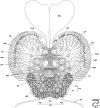
Fig 5. Anterior brain organization of Riftia.
Scheme of histological cross section based on the anterior brain sections of a 79-mm-long specimen (S1 Fig). Level of section shown at diagram at bottom right. The enteral coelom (EC) enables demarcating position of supra- and subesophageal elements. Enteral coelom (EC) overlain by structures of supraesophageal ganglion: peripheral perikarya (nep) surround neuropile of lateral brain lobes (NE). Pair of NE connected via dorsal commissure (DC). Obturacular neurite bundles (OBN) enter DC. Structures of subesophageal ganglion (tripartite ventral aggregation of perikarya) underlie enteral coelom (EC). amp–anterior median aggregation of perikarya, DC–dorsal commissure, DLN–dorsal longitudinal bundles, dop–dorsal aggregation of perikarya, EP–epidermis, EC–enteral coelom, LNT–longitudinal nerve tracts projecting from VNC into brain, LR–undifferential tentacle lamellae, NE–neuropile of lateral brain lobes, nep–peripheral perikarya of lateral brain lobes, OBC–obturacular coelom, OBL–obturacular lobes, OBN–obturacular neurite bundles, OBV–obturacular blood vessels, SLN–supraenteral longitudinal neurite bundles, TEN–neurite bundles of tentacles (palps), VSN–vertical supraenteral neurite bundles, vtp—tripartite ventral aggregation of perikarya, vvtp–ventral perikarya of vtp, XXL–pair of prominent bundles of large longitudinal nerve tracts (part of LNT).
Fig 6
Fig 6. Middle brain organization of Riftia.
Scheme of cross section based on the midbrain histological sections of a 79-mm-long specimen (S2 Fig). Level of section shown at diagram at bottom right. Midbrain’s elements of supraesophageal ganglion: neuropile of lateral brain lobes (NE) connected via dorsal commissure (DC) and supraenteral commissure (SPC). Midbrain’s elements of subesophageal ganglion: neuropile of tripartite ventral aggregation of perikarya (vtp) connected via subenteral commissure (SBC). Longitudinal nerve tracts (LNT), as circumesophageal connectives, surround enteral coelom (EC). In dorsal brain part they give rise to DC, SPC, whereas in ventral brain they are connected via SBC. DC–dorsal commissure, DLN–dorsal longitudinal bundles, dop–dorsal aggregation of perikarya, EP–epidermis, GA–giant axons, EC–enteral coelom, ECM–extracellular matrix, LNT–longitudinal nerve tracts projecting from VNC into brain, LR–undifferential tentacle lamellae, lvtp–ventrolateral perikarya of vtp, NE–neuropile of lateral brain lobes, nep–peripheral perikarya of lateral brain lobes, OBC–obturacular coelom, OBL–obturacular lobes, OBV–obturacular blood vessels, pl–large perikarya, ps–small perikarya, SBC–subenteral commissure, SPC–supraenteral commissure, TEN–neurite bundles of tentacles (palps), vtp–tripartite ventral aggregation of perikarya, VPN–posterior vertical median bundles.
Fig 7
Fig 7. Posterior brain organization of Riftia.
Schematic cross section based on the posterior brain histological sections of a 79-mm-long specimen (S3 Fig). Level of section shown at diagram at bottom right. Posterior brain elements of supraesophageal ganglion: neuropile of lateral brain lobes (NE) connected via dorsal commissure (DC). Posterior brain elements of subesophageal ganglion: neuropile of tripartite ventral aggregation of perikarya (vtp) connected via subenteral commissure (SBC). Longitudinal nerve tracts (LNT) project from ventral nerve cord (VNC) into brain. DC–dorsal commissure, ECM–extracellular matrix, EP–epidermis, GA–giant axons, EC–enteral coelom, LNT–longitudinal nerve tracts projecting from ventral nerve cord into brain, LR–undifferential tentacle lamellae, lvtp–ventrolateral perikarya of vtp, NE–neuropile of lateral brain lobes, nep–peripheral perikarya of lateral brain lobes, OBC–obturacular coelom, OBV–obturacular blood vessels, pl–large perikarya, pmp–posterior median perikarya aggregation, ps–small perikarya, SBC–subenteral commissure, TEN–neurite bundles of tentacles (palps), vtp–tripartite ventral aggregation of perikarya, vvtp–ventral perikarya of vtp.
Fig 8
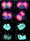
Fig 8. Supra- and subesophageal ganglia in Riftia.
3D models of the Riftia brain. Enteral coelom (EC) containing the gut demarcates the dorsal and ventral parts of the brain. Dorsal part homologous to supraesophageal ganglion, ventral part to subesophageal ganglion. A-D–supraesophageal neuronal elements. E-H–subesophageal neuronal elements in Riftia brain. View sides shown at bottom right of each image. Cube side 255 μm. Dashed lines: neural elements under transparent structures. amp–anterior median aggregation of perikarya, DC–dorsal commissure, DLN–dorsal longitudinal bundles, GA–giant axons, gap–giant perikarya, EC–enteral coelom, LNT–longitudinal nerve tracts projecting from ventral nerve cord into brain, lvtp–ventrolateral perikarya of vtp, NE–neuropile of lateral brain lobes, nep–peripheral perikarya of lateral brain lobes, pmp–posterior median perikarya aggregation, SBC–subenteral commissure, SPC–supraenteral commissure, SLN–supraenteral longitudinal neurite bundles, VPN–posterior vertical median bundles, VSN–vertical supraenteral neurite bundles, vtp–tripartite ventral aggregation of perikarya, vvtp–ventral perikarya of vtp.
Fig 9
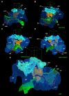
Fig 9. Longitudinal nerve tracts and main commissures in Riftia brain.
3D models of the Riftia brain. Longitudinal nerve tracts (LNT), as circumesophageal connectives, connect supra- and subesophageal ganglia and give rise to dorsal commissure (DC) and supraenteral commissure (SPC) in supraesophageal ganglion, and to subenteral commissure (SBC) in subesophageal ganglion. Giant axons running from giant perikaya lie in anterior DC (not shown) to ventral nerve cord. A-E–main commissures (dorsal, DC, supra-, SPC, and subesophageal, SBC) and longitudinal nerve tracts (LNT). The latter is homologous to circumesophageal connectives in other annelid brains. View sides shown at bottom right of each image. Cube side 255 μm. Dashed lines: neural elements under transparent structures. DC–dorsal commissure, GA–giant axons, EC–enteral coelom, LNT–longitudinal nerve tracts projecting from ventral nerve cord into brain, SBC–subenteral commissure, SPC–supraenteral commissure, XXL–pair of prominent bundles of large longitudinal nerve tracts (part of LNT).
Fig 10
Fig 10. Histological details in the anterior brain of Riftia.
In the anterior brain, the dorsal commissure gives rise to the obturacular neurite bundles (OBN) (A). Giant axons (GA) extend from giant perikarya (gap) and follow the anterior vertical median bundles (VAN), which cross each other (B-D). Anterior and posterior vertical median bundles (VAN, VPN) are present. The latter do not cross each other (D, E). Thick cuticle layer (cuticular shield, CUP) protects apical surface of brain (G). A–obturacular neurite bundles (OBN) connecting with dorsal commissure (DC). B, C–giant perikarya with clear nuclei in juvenile brain. D–anterior vertical median bundles (VAN) composed of giant axons (GA). E–posterior vertical median bundles (VPN) with no giant axon. F–giant perikarion degrading in brain of 79-mm-long male. G–cuticular plate protecting brain (CUP). ECM–extracellular matrix, CU–cuticle, CUP–cuticle schield, DC–dorsal commissure, DLN–dorsal longitudinal bundles, dop–dorsal aggregation of perikarya, EP–epidermis, GA–giant axons, gap–giant perikarya, LNT–longitudinal nerve tracts projecting from VNC into brain, NE–neuropile of lateral brain lobes, nep–peripheral perikarya of lateral brain lobes, OBC–obturacular coelom, OBL–obturacular lobes, OBN–obturacular neurite bundles, OBV–obturacular blood vessels, pl–large perikarya, ps–small perikarya, SPC–supraenteral commissure, TEN–neurite bundles of tentacles (palps), vtp–tripartite ventral aggregation of perikarya, VPN–posterior vertical median bundles, VAN–anterior vertical median bundles, XXL–pair of prominent bundles of large longitudinal nerve tracts (part of LNT).
Fig 11
Fig 11. Hypothetical vestimentiferan brain origin.
Orrhage and Müller [47] showed the general presence of 4 transverse commissures in the brain (a.k.a. supraesophageal ganglion) of numerous annelids (A), which join through their roots and circumesophageal connectives to the ventral nerve cords. The palp neurites extend from the dorsal root of the circumesophageal connectives in annelids. In vestimentiferans (C), the “brain” is a fusion of the supra- and subesophageal ganglia. Longitudinal nerve tracts (LNT), as circumesophageal connectives, connect the ganglia. The dorsal (DC) and supraenteral (SPC) commissures connect transversely the paired structures of the supraesophageal ganglion, whereas the subenteral commissure (SBC) connects transversely the structures of the subesophageal ganglion. The innervation of tentacles from the DC makes them homologous to peristomial palps of other annelids. Hypothetical ancestral state of vestimentiferan brain (B) is transitional between annelid brain (A) and vestimentiferan brain (C). Dorsal side to the top. A–supra- and subesophageal ganglia in annelids (after [47]). B–hypothetical transitional state. C–vestimentiferan brain. APN–neurite bundles of palps, B–brain, C–commissure of sbg, CC–circumesophageal connectives, DC–dorsal commissure, DRCC–dorsal (posterior) root of the CC, EC–enteral coelom, LNT–longitudinal nerve tracts projecting from ventral nerve cord into brain, sbg–subesophageal ganglion, SBC–subenteral commissure, spg–supraesophageal ganglion (the brain in annelids), SPC–supraenteral commissure, VRCC–ventral (anterior) root of CC, TEN–neurite bundles of tentacles (palps).
Fig 12
Fig 12. Reconstruction of hypothesized neural ancestor traits of the siboglinid central nervous system.
Main neural characters of siboglinids and annelid sister groups used to reconstruct neural archetype of siboglinid ancestor. For neural characters to homologize CNS in siboglinids and annelid sister groups, see table (A) and diagrams (B-K). In table (A), “?” means “not known because not studied in detail”. We suggest the neural archetype of the siboglinid ancestor to have had separate supraesophageal (= brain) and subesophageal ganglia, two commissures of circumesophageal connectives which were interconnected by at least one pair of crossing neurites and possibly gave rise to numerous neurites into palps. Giant perikarya are places in the supraesophageal ganglion. Our hypothesis: the VNC of the siboglinid ancestor was paired (or at least paired in the anterior part) and non-ganglionated, with a pair of giant axons. Relation among siboglinid groups based on a combination of recent phylogenetic trees: frenulate clade resolved based on cladistic analysis [7], interrelationship of siboglinid clades on phylogenetic and phylogenomic data [6,85, 86, 87], annelid outgroups on phylogenomic data [13]. Neural diagrams include left (upper row) and dorsal views (lower row) of the anteriormost body. Anterior/ dorsalside are at top. Perikarya position shown in grey. Cerebral elements drawn based on larvae of vestimentiferan Riftia pachyptila [25] (B); adult Riftia pachyptila (this study) (C); Osedax knutei (= O. “nudepalp E” [35,85]) (D); frenulates Polybrachia annulata (E) Siboglinum caulleryi [27,28] (F) and Nereilinum murmanicum [31] (G), cirratulid Cirratulus cf. cirratus [51,68,88] (I), sabellid Euchone papillosa [49,68] (J), and spionid Malacoceros fuliginosus [47,50,52,53,64,68] (K). Reconstructed archetype H1 has a combination of the hypothesized ancestral siboglinid states (H). Dashed lines: hypothesized neuronal elements in siboglinid ancestor. ACBR–anterior commissure of brain, ADN–antero-dorsal nerve, aga–ganglion-like aggregation at base of TEC, ANN–anterior nerve net, B–brain, C–commissure in sbg, CC–circumesophageal connectives, ccl–commissural cell cluster; COM–commissure, CON–connective, DC–dorsal commissure, DRCC–dorsal (posterior) root of the CC, EC–enteral coelom, LLN–lateral longitudinal neurite bundles in brain, LNT–longitudinal nerve tracts projecting from VNC into brain, MCC–middle cross commissure, NA–nerve ring (after [28]) or brain ring (after [31]), OBN–obturacular neurites, PCBR–posterior commissure of brain, PN–palp nerve, TEC–tentacular commissure, TEN–neurite bundles of tentacles (palps), SBC–subenteral commissure, sbg–subesophageal ganglion, SPC–supraenteral commissure, spg–supraesophageal ganglion, VAN–anterior vertical median bundles, VNC–ventral nerve cord, VRCC–ventral (anterior) root of CC.

Similar articles

Cited by

References

    1. Schulze A, Halanych KM. Siboglinid evolution shaped by habitat preference and sulfide tolerance. Hydrobiologia. 2003;496: 199–205. 10.1023/A:1026192715095 - DOI
    1. Halanych KM. Molecular phylogeny of siboglinid annelids (a.k.a. pogonophorans): a review. Hydrobiologia. 2005;535–536: 297–307. 10.1007/s10750-004-1437-6 - DOI
    1. Karaseva NP, Rimskaya-Korsakova NN, Galkin SV, Malakhov VV. Taxonomy, geographical and bathymetric distribution of vestimentiferan tubeworms (Annelida, Siboglinidae). Biol Bull. 2016;43 10.1134/S1062359016090132 - DOI
    1. Webb M. Lamellibrachia barhami, Gen. Nov., Sp. Nov., (Pogonophora), from the Northeast Pacific. Bull Mar Sci. 1969;19: 18–47.
    1. Southward EC. Siboga-Expeditie Pogonophora. Siboga-Expeditie Uitkomsten op Zool Bonatisch, Oceanogr en Geol Geb verzameld Ned Oost-Indië 1899–1900. 1961;25: 1–22.

Publication types