

Next-generation sequencing identifies unexpected genotype-phenotype correlations in patients with retinitis pigmentosa
- PMID: 30543658
- PMCID: PMC6292620
- DOI: 10.1371/journal.pone.0207958
Next-generation sequencing identifies unexpected genotype-phenotype correlations in patients with retinitis pigmentosa
Abstract
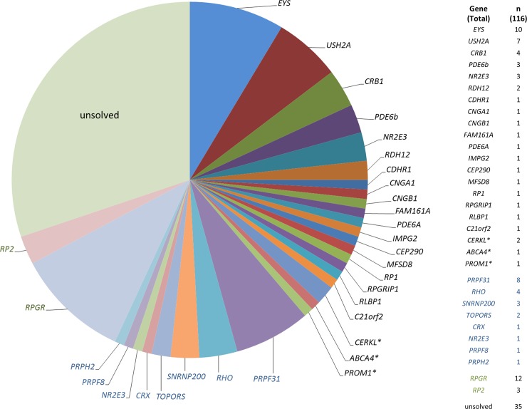
Retinitis pigmentosa (RP) is an inherited degenerative disease causing severe retinal dystrophy and visual impairment mainly with onset in infancy or adolescence. Targeted next-generation sequencing (NGS) has become an efficient tool to encounter the enormous genetic heterogeneity of diverse retinal dystrophies, including RP. To identify disease-causing mutations in unselected, consecutive RP patients, we conducted Sanger sequencing of genes commonly involved in the suspected genetic RP subtype, followed by targeted large-panel NGS if no mutation was identified, or NGS as primary analysis. A high (70%) detection rate of disease-causing mutations was achieved in a large cohort of 116 unrelated patients. About half (48%) of the solved RP cases were explained by mutations in four genes: RPGR, EYS, PRPF31 and USH2A. Overall, 110 different mutations distributed across 30 different genes were detected, and 46 of these mutations were novel. A molecular diagnosis was achieved in the majority (82-100%) of patients if the family history was suggestive for a particular mode of inheritance, but only in 60% in cases of sporadic RP. The diagnostic potential of extensive molecular analysis in a routine setting is also illustrated by the identification of unexpected genotype-phenotype correlations for RP patients with mutations in CRX, CEP290, RPGRIP1, MFSD8. Furthermore, we identified numerous mutations in autosomal dominant (PRPF31, PRPH2, CRX) and X-linked (RPGR) RP genes in patients with sporadic RP. Variants in RP2 and RPGR were also found in female RP patients with apparently sporadic or dominant disease. In summary, this study demonstrates that massively parallel sequencing of all known retinal dystrophy genes is a valuable diagnostic approach for RP patients.
Conflict of interest statement
I have read the journal's policy and the authors of this manuscript have the following competing interests: Dres. Neuhaus, Lenzner, Zahnleiter, Betz, and Eisenberger are employees of Bioscientia, a publicly traded diagnostic company. Dr. Bolz has been employee of Bioscientia until 2016. There are no patents, products in development or marketed products to declare. This does not alter the authors' adherence to PLOS ONE policies on sharing data and materials. The other authors have declared that no competing interests exist.
Figures

Similar articles
-
Next-generation sequencing to solve complex inherited retinal dystrophy: A case series of multiple genes contributing to disease in extended families.Mol Vis. 2017 Jul 20;23:470-481. eCollection 2017. Mol Vis. 2017. PMID: 28761320 Free PMC article.
-
Targeted Next-Generation Sequencing Improves the Diagnosis of Autosomal Dominant Retinitis Pigmentosa in Spanish Patients.Invest Ophthalmol Vis Sci. 2015 Apr;56(4):2173-82. doi: 10.1167/iovs.14-16178. Invest Ophthalmol Vis Sci. 2015. PMID: 25698705
-
Comprehensive survey of mutations in RP2 and RPGR in patients affected with distinct retinal dystrophies: genotype-phenotype correlations and impact on genetic counseling.Hum Mutat. 2007 Jan;28(1):81-91. doi: 10.1002/humu.20417. Hum Mutat. 2007. PMID: 16969763
-
[Genetics of retinitis pigmentosa: metabolic classification and phenotype/genotype correlations].J Fr Ophtalmol. 2005 Jan;28(1):71-92. doi: 10.1016/s0181-5512(05)81029-0. J Fr Ophtalmol. 2005. PMID: 15767903 Review. French.
-
Genes and mutations causing retinitis pigmentosa.Clin Genet. 2013 Aug;84(2):132-41. doi: 10.1111/cge.12203. Epub 2013 Jun 19. Clin Genet. 2013. PMID: 23701314 Free PMC article. Review.
Cited by
-
Mutation spectrum of PRPF31, genotype-phenotype correlation in retinitis pigmentosa, and opportunities for therapy.Exp Eye Res. 2020 Mar;192:107950. doi: 10.1016/j.exer.2020.107950. Epub 2020 Jan 31. Exp Eye Res. 2020. PMID: 32014492 Free PMC article. Review.
-
A Novel Arg120Pro Mutation in the RP2 Gene in an Iranian Family with X-linked Retinitis Pigmentosa: A Case Report.Iran J Med Sci. 2023 Nov 1;48(6):606-611. doi: 10.30476/IJMS.2022.96392.2792. eCollection 2023 Nov. Iran J Med Sci. 2023. PMID: 38094283 Free PMC article.
-
Autosomal Dominant Retinitis Pigmentosa-Associated TOPORS Protein Truncating Variants Are Exclusively Located in the Region of Amino Acid Residues 807 to 867.Invest Ophthalmol Vis Sci. 2022 May 2;63(5):19. doi: 10.1167/iovs.63.5.19. Invest Ophthalmol Vis Sci. 2022. PMID: 35579903 Free PMC article.
-
Neuronal Ceroid Lipofuscinoses Type 7 (CLN7)- A Case Series Reporting Cross Sectional and Retrospective Clinical Data to Evaluate Validity of Standardized Tools to Assess Disease Progression, Quality of Life, and Adaptive Skills.Res Sq [Preprint]. 2024 Jun 26:rs.3.rs-3983366. doi: 10.21203/rs.3.rs-3983366/v1. Res Sq. 2024. Update in: Orphanet J Rare Dis. 2024 Dec 19;19(1):468. doi: 10.1186/s13023-024-03448-8. PMID: 38978590 Free PMC article. Updated. Preprint.
-
State of the Art on Inherited Retinal Dystrophies: Management and Molecular Genetics.J Clin Med. 2025 May 18;14(10):3526. doi: 10.3390/jcm14103526. J Clin Med. 2025. PMID: 40429522 Free PMC article. Review.
References
-
- Hamel C. Retinitis pigmentosa. Orphanet J Rare Dis. 2006;1:40 Epub 2006/10/13. 10.1186/1750-1172-1-40 ; PubMed Central PMCID: PMC1621055. - DOI - PMC - PubMed
-
- Hartong DT, Berson EL, Dryja TP. Retinitis pigmentosa. Lancet. 2006;368(9549):1795–809. Epub 2006/11/23. 10.1016/S0140-6736(06)69740-7 . - DOI - PubMed
-
- Martinez-Fernandez De La Camara C, Nanda A, Salvetti AP, Fischer MD, MacLaren RE. Gene therapy for the treatment of X-linked retinitis pigmentosa. Expert Opin Orphan Drugs. 2018;6(3):167–77. Epub 2018/07/31. 10.1080/21678707.2018.1444476 ; PubMed Central PMCID: PMC6059358. - DOI - PMC - PubMed
-
- Neveling K, Collin RW, Gilissen C, van Huet RA, Visser L, Kwint MP, et al. Next-generation genetic testing for retinitis pigmentosa. Hum Mutat. 2012;33(6):963–72. Epub 2012/02/16. 10.1002/humu.22045 ; PubMed Central PMCID: PMC3490376. - DOI - PMC - PubMed
-
- O'Sullivan J, Mullaney BG, Bhaskar SS, Dickerson JE, Hall G, O'Grady A, et al. A paradigm shift in the delivery of services for diagnosis of inherited retinal disease. J Med Genet. 2012;49(5):322–6. Epub 2012/05/15. 10.1136/jmedgenet-2012-100847 . - DOI - PubMed
Publication types
MeSH terms
LinkOut - more resources
Full Text Sources
Medical
