Case Reports
. 2018 Dec 13;14(12):e1007845.
doi: 10.1371/journal.pgen.1007845. eCollection 2018 Dec.

Biallelic mutations in nucleoporin NUP88 cause lethal fetal akinesia deformation sequence

Affiliations
Case Reports

Biallelic mutations in nucleoporin NUP88 cause lethal fetal akinesia deformation sequence

Edith Bonnin et al. PLoS Genet. .

Abstract

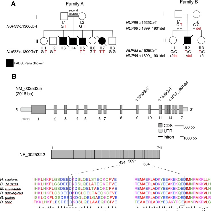
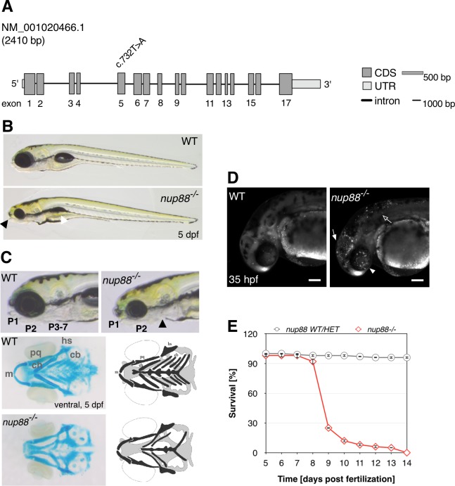
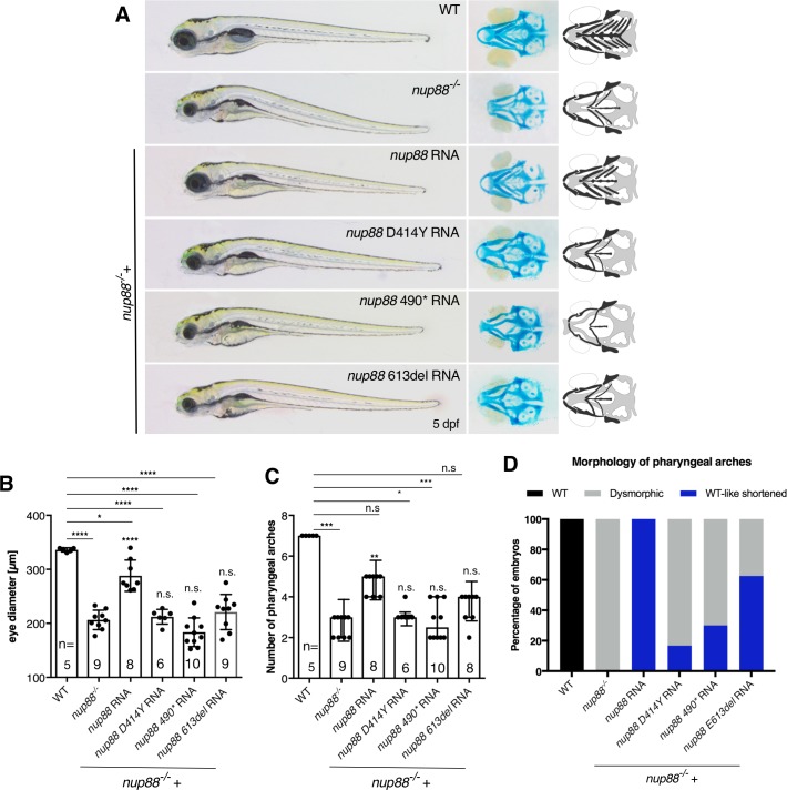
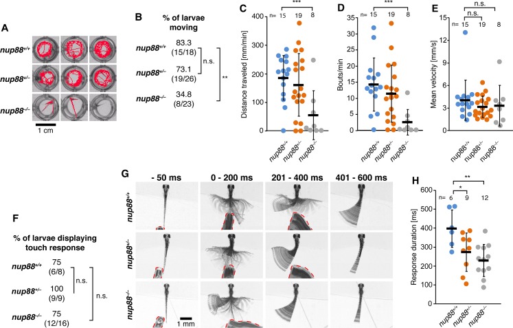
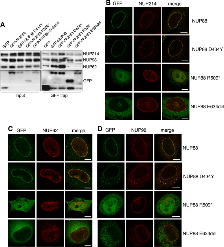
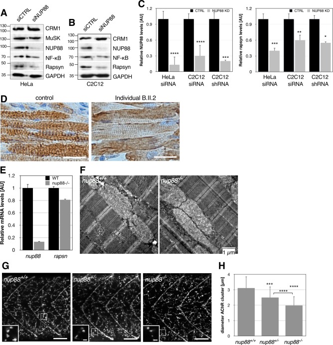
Nucleoporins build the nuclear pore complex (NPC), which, as sole gate for nuclear-cytoplasmic exchange, is of outmost importance for normal cell function. Defects in the process of nucleocytoplasmic transport or in its machinery have been frequently described in human diseases, such as cancer and neurodegenerative disorders, but only in a few cases of developmental disorders. Here we report biallelic mutations in the nucleoporin NUP88 as a novel cause of lethal fetal akinesia deformation sequence (FADS) in two families. FADS comprises a spectrum of clinically and genetically heterogeneous disorders with congenital malformations related to impaired fetal movement. We show that genetic disruption of nup88 in zebrafish results in pleiotropic developmental defects reminiscent of those seen in affected human fetuses, including locomotor defects as well as defects at neuromuscular junctions. Phenotypic alterations become visible at distinct developmental stages, both in affected human fetuses and in zebrafish, whereas early stages of development are apparently normal. The zebrafish phenotypes caused by nup88 deficiency are rescued by expressing wild-type Nup88 but not the disease-linked mutant forms of Nup88. Furthermore, using human and mouse cell lines as well as immunohistochemistry on fetal muscle tissue, we demonstrate that NUP88 depletion affects rapsyn, a key regulator of the muscle nicotinic acetylcholine receptor at the neuromuscular junction. Together, our studies provide the first characterization of NUP88 in vertebrate development, expand our understanding of the molecular events causing FADS, and suggest that variants in NUP88 should be investigated in cases of FADS.


Conflict of interest statement

The authors have declared that no competing interests exist.

Figures

Fig 1
Fig 1. NUP88 mutations identified in affected individuals from two families.
(A) Pedigrees of two families identified with mutations in NUP88 (GenBank: NM_002532.5). (B) NUP88 gene and protein structure, location of the identified mutations, and phylogenetic conservation of the mutated residues and surrounding amino acids. Identical amino acids are indicated by asterisks, highly similar residues by colons.
Fig 2
Fig 2. Modelling of human NUP88.
(A) The N-terminal domain of NUP88 reveals a seven-bladed ß-propeller with an N-terminal extension. The rainbow coloring indicates N-terminal residues in blue (NTD = N-terminal domain) and C-terminal residues of the propeller in red. Individual blades are indicated by numbers. The red arrow indicates the location of the p.D434Y point mutation (see below for details). (B) Overlay of the NUP88 model (red) with the X-ray structures of Nup82 from baker’s yeast (PDBid: 3pbp, in gray) and C. thermophilum (PDBid: 5cww; in light yellow). Significant differences between species are in blade 4 (HTH-motif; black arrow) and 5 (extended loop; blue arrow). (C) Composite model of the N- and C-terminal regions of NUP88. The presented model was generated using RaptorX with its standard settings and misses about 40 amino acid residues after the propeller region. Both, the propeller and CTD regions are colored in rainbow coloring as in (A). The individual mutations are indicated by their numbering and represented in sphere mode. (D) Magnification of the loop bearing the D434 mutation in NUP88 in stick mode. The coloring of the individual molecules is as described in (B).
Fig 3
Fig 3. Morphological phenotypes of nup88-/- mutants.
(A) nup88 gene structure and location of the mutation in the sa2206 allele. (B) Lateral view of wild-type and nup88-/- embryos at 5 dpf. nup88-/- mutants resemble the flathead group of mutants characterized by decreased head and eye size and the absence of a protruding mouth (black arrowhead). The larvae furthermore show aplastic swim bladder (white arrowhead), hypoplastic liver, abnormal gut and a marked curvature of the anterio-posterior axis. (C) Higher magnification lateral views of the head region of wild-type and nup88-/- embryos at 5 dpf. Alcian blue staining of the viscerocranium revealed that nup88-/- mutants lack pharyngeal arches 3 to 7 (P3-7). A ventral view of the head showed that hyoid and mandibular arches (P1 and P2) were present, but dysmorphic. A schematic representation of the viscerocranium is shown to illustrate alcian-blue images. Genotypes of larvae were determined by fin clip and RFLP. m, Meckel's cartilage; ch, ceratohyal; pq, palatoquadrate; cb, ceratobranchials (P3-7); hs, hyosymplectic. (D) Acridine orange staining revealed an increase in apoptotic cells in the head, including the eyes (arrowhead), the brain (filled arrow), and anterior part of the trunk (arrow) of nup88-/- mutants at 35 hpf compared with wild-type siblings. Shown are confocal images. Scale bars, 100 μm. (E) Survival curves of nup88 mutants and siblings. 75 larvae were analyzed in each category. Error bars are ±SEM.
Fig 4
Fig 4. Wild-type nup88, but not disease-related mutant forms, rescue defects of nup88-/- embryos.
(A) nup88+/- embryos were in-crossed and 300 pg of mRNA encoding wild-type or the respective mutant nup88 were microinjected at the one-cell stage. The extent of rescue of each variant mRNA was evaluated after 5 dpf using (B) the diameter of the eye (One-way ANOVA test), (C) the number of pharyngeal arches (Kruskal-Wallis test) and (D) their morphology as readouts. Only injecting wild-type, but not the mutant forms of nup88 mRNA, rescued the reduced eye size and the number of pharyngeal arches as revealed by Alcian blue staining (A). At least three independent injections were performed for each condition. Values are mean ± SD. Significance in comparison to nup88+/+ and nup88-/- embryos, respectively. n.s., non- significant, *P<0.05, **P<0.01, ***P<0.001, ****P<0.0001. The statistical annotations appearing just above the histogram bars refer to the comparison to the uninjected mutants. n is number of embryos/larvae analyzed. In (D) 21 larvae from 3 independent experiments (3x7) were analyzed for each form.
Fig 5
Fig 5. nup88 mutants display motor impairments.
(A) Images showing representative examples of motion tracking (red lines) of 4 dpf nup88 homozygous and heterozygous mutants, and wild-type controls during one-minute long spontaneous locomotion recordings. (B) Quantification of percentages of larvae displaying spontaneous movement, also shown as (number of moving larvae/total number of larvae). Beeswarm graphs with individual data points depicting distance travelled/minute (C), bouts/minute (D), and mean velocity of swimming bouts (E) of nup88 mutants and wild-type controls displaying spontaneous locomotion. (F) Quantification of percentages of larvae displaying touch-induced escape response, also shown as (number of responsive larvae/total number of larvae). (G) Representative examples of touch-induced escape behavior of 4 dpf head-restrained nup88 mutant and wild-type larvae. The first column depicts single frames taken 50 ms before an escape response was induced by touching the trunk of larvae with a pipette tip (red dashed lines). The other columns show superimposed frames of escape responses at consecutive time intervals after touch. The asymmetric tail movement displayed in some of the images (nup88+/- and nup88-/- at 201–400 ms, and all genotypes at 401–600 ms) represents slow motion of the tail toward its original position. The escape response terminated before this phase (see also S2–S4 Movies). (H) Graph showing average durations of escape response in nup88 mutants and wild-types. * p < 0.05, ** p < 0.004, *** p = 0.001, n.s. = non-significant, two-tailed Fisher exact test (B and G) or two-tailed t-test (C-E, H). Data in C-E and H are shown as mean ± SD. n is number of embryos/larvae analyzed.
Fig 6
Fig 6. NUP88 mutants have distinct effects on NUP88 nucleoporin partners.
(A) GFP-trap assays to study the effects of NUP88 mutants on its interaction with NUP214, NUP62 and NUP98. GFP and GFP-NUP88 fusion proteins were transiently expressed in HeLa cells. After 48 h, GFP proteins and associated factors were recovered from cell lysates and probed by Western blot analysis using antibodies against Nup214, NUP62, and NUP98. Successful transfection and expression of the proteins was confirmed by probing with antibodies against GFP. GFP proteins had the following size: GFP-NUP88 WT, D434Y, and E634del: ~115 (88 + 27) kDa; GFP-NUP88 R509*: 83 (56 + 27) kDa; GFP: 27 kDa. (B-D) HeLa cells were transiently transfected with GFP-constructs of the respective NUP88 mutant and fixed and stained after 48 h for immunofluorescence microscopy using (B) anti-NUP214, (C) anti-NUP62, and (D) anti-NUP98 antibodies. Only NUP88 E634del affects NUP62 association with NPCs. Scale bars: 10 μm.
Fig 7
Fig 7. Loss of functional NUP88 affects rapsyn expression as well as AChR clustering.
(A) HeLa and (B) C2C12 cells were treated with the indicated siRNAs for 2 days and cellular lysates were subjected to Western blot analysis using antibodies against NUP88, CRM1, MuSK, NF-kB, and rapsyn. GAPDH was used as loading control. Rapsyn protein levels are reduced in NUP88-depleted cells. Note MuSK has a predicted molecular weight of 97 kDa, but migrates higher [20]. (C) Quantification of the respective expression levels of NUP88 and rapsyn after transfection of HeLa and C2C12 cells with the indicated siRNAs and shRNA-mediated depletion of NUP88 in C2C12 cells. Blots from three independent experiments for each condition were analyzed. Data present mean ± SEM. P-values ****<0.0001, ***<0.001; **<0.01, *<0.05; t-test, one-tailed. (D) Bright-field images of histological muscle sections from individual B.II.2 and a control fetus stained with anti-rapsyn antibodies (brown). Nuclei were visualized by hematoxylin. (E) qRT-PCR analysis of nup88 and rapsn transcripts in 5 dpf wild-type and nup88-/- larvae. (F) Skeletal muscle organization remains unaffected in nup88-/- zebrafish larvae. Larvae were prepared at 5 dpf for transmission electron microscopy. Skeletal muscle of WT and mutant zebrafish show intact myofibril alignment with their regularly stacked Z-line and clearly identifiable H- and I-zones. Shown are representative longitudinal sections of skeletal muscle from nup88+/+ and nup88-/- larvae, respectively. Scale bar, 1 μm. (G) Anterior trunk regions of wild-type (left) and nup88 heterozygous and homozygous mutant larvae were stained with antibodies against the AChR and secondary Alexa 488 antibodies. AChR cluster size was significantly reduced in nup88-/- larvae as compared to nup88+/+ and nup88+/- larvae (higher magnification insets). Insets are taken from the marked area in the respective overview image. Inset for nup88-/- was rotated by 180°. Scale bars, 50 μm (overview), 5 μm (insets). (H) Quantification of the AChR size in 4 dpf nup88+/+ (n = 8), nup88+/- (n = 14), and nup88-/- (n = 12) larvae. Per larvae 100 clusters throughout z-stacks of confocal images were manually measured using ImageJ. Data present mean ± SD. P-values ****<0.0001, ***<0.001; t-test, one-tailed.

Similar articles

Cited by

References

    1. Dickmanns A, Kehlenbach RH, Fahrenkrog B. Nuclear Pore Complexes and Nucleocytoplasmic Transport: From Structure to Function to Disease. Int Rev Cell Mol Biol. 2015;320:171–233. 10.1016/bs.ircmb.2015.07.010 . - DOI - PubMed
    1. Lussi YC, Hugi I, Laurell E, Kutay U, Fahrenkrog B. The nucleoporin Nup88 is interacting with nuclear lamin A. Mol Biol Cell. 2011;22(7):1080–90. Epub 2011/02/04. doi: mbc.E10-05-0463 [pii] 10.1091/mbc.E10-05-0463 ; PubMed Central PMCID: PMC3069011. - DOI - PMC - PubMed
    1. Fornerod M, van Deursen J, van Baal S, Reynolds A, Davis D, Murti KG, et al. The human homologue of yeast CRM1 is in a dynamic subcomplex with CAN/Nup214 and a novel nuclear pore component Nup88. Embo J. 1997;16(4):807–16. Epub 1997/02/17. 10.1093/emboj/16.4.807 ; PubMed Central PMCID: PMC1169681. - DOI - PMC - PubMed
    1. Bastos R, Ribas de Pouplana L, Enarson M, Bodoor K, Burke B. Nup84, a novel nucleoporin that is associated with CAN/Nup214 on the cytoplasmic face of the nuclear pore complex. J Cell Biol. 1997;137(5):989–1000. Epub 1997/06/02. ; PubMed Central PMCID: PMC2136229. - PMC - PubMed
    1. Griffis ER, Xu S, Powers MA. Nup98 localizes to both nuclear and cytoplasmic sides of the nuclear pore and binds to two distinct nucleoporin subcomplexes. Mol Biol Cell. 2003;14(2):600–10. 10.1091/mbc.E02-09-0582 . - DOI - PMC - PubMed

Publication types

MeSH terms

Supplementary concepts