. 2018 Dec 13;13(12):e0208636.
doi: 10.1371/journal.pone.0208636. eCollection 2018.

Epitope specificity of anti-synapsin autoantibodies: Differential targeting of synapsin I domains

Affiliations

Epitope specificity of anti-synapsin autoantibodies: Differential targeting of synapsin I domains

Robert Mertens et al. PLoS One. .

Abstract

Objective: To identify the specific domains of the presynaptic protein synapsin targeted by recently described autoantibodies to synapsin.

Methods: Sera of 20 and CSF of two patients with different psychiatric and neurological disorders previously tested positive for immunoglobulin (Ig)G antibodies to full-length synapsin were screened for IgG against synapsin I domains using HEK293 cells transfected with constructs encoding different domains of rat synapsin Ia. Additionally, IgG subclasses were determined using full-length synapsin Ia. Serum and CSF from one patient were also screened for IgA autoantibodies to synapsin I domains. Sera from nine and CSF from two healthy subjects were analyzed as controls.

Results: IgG in serum from 12 of 20 IgG synapsin full-length positive patients, but from none of the healthy controls, bound to synapsin domains. Of these 12 sera, six bound to the A domain, five to the D domain, and one to the B- (and possibly A-), D-, and E-domains of synapsin I. IgG antibodies to the D-domain were also detected in one of the CSF samples. Determination of IgG subclasses detected IgG1 in two sera and one CSF, IgG2 in none of the samples, IgG3 in two sera, and IgG4 in eight sera. One patient known to be positive for IgA antibodies to full-length synapsin had IgA antibodies to the D-domain in serum and CSF.

Conclusions: Anti-synapsin autoantibodies preferentially bind to either the A- or the D-domain of synapsin I.


Conflict of interest statement

The authors have declared that no competing interests exist.

Figures

Fig 1
Fig 1. Specific recognition of synapsin I domains expressed in transfected HEK cells by domain-specific antibodies.
(A) Domain structure of the synapsin isoforms Ia and Ib. The N-terminal domains A-C are highly conserved in all synapsins. The C-terminal region is variable due to heterogeneous combinations of domains. (B) Diagram of the synapsin Ia fragments examined to investigate targeting of patient synapsin autoantibodies. (C-H) HEK293 cells were transfected with rat synapsin Ia constructs, comprising the eYFP-tagged fragment ABC (C), eGFP-tagged BC (D), eGFP-tagged C (E), eGFP-tagged DE (F), eGFP-tagged D (G), or eGFP-tagged E domain (H). Cells were fixed, permeabilized and incubated with antibodies either directed against the A-domain of synapsin I and II (rabbit anti-synapsin A; dilution 1:500), the D-domain of synapsin I (mouse anti-synapsin D; dilution 1:500), the C-domain of synapsin I/II (rabbit anti-synapsin C; dilution 1:500), or the E-domain of synapsin Ia (rabbit anti-synapsin E; dilution 1:200). Antibody binding was visualized using Alexa Fluor 549-coupled secondary antibodies. Transfected cells were correctly recognized by the respective antibodies that colocalized with the signal of the tags, confirming domain identities and detectability of the various domains by anti-synapsin antibodies.
Fig 2
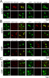
Fig 2. Serum IgG autoantibodies against the A-domain of a patient with bipolar affective disorder and serum and CSF IgG autoantibodies against the D-domain of a patient with a clinically isolated syndrome (CIS).
(A) HEK293 cells were transfected with eYFP-tagged synapsin I fragment ABC, eGFP-tagged DE, BC or C. Cells were incubated with patient serum at a dilution of 1:320. Bound IgG was detected using an Alexa 594-coupled secondary antibody to the Fc fragment of human IgG. The patient’s serum demonstrated positive immunoreactivity that co-localized with the eYFP tag of synapsin ABC, but with none of the other constructs. (B) HEK293 cells were transfected with eYFP-tagged synapsin I fragment ABC, eGFP-tagged DE, D or E. Cells were incubated with patient serum or CSF at a dilution of 1:320. Bound IgG was detected using an Alexa 594-coupled secondary antibody to the Fc fragment of human IgG. Both the patient’s serum and CSF demonstrated positive immunoreactivity that co-localized with the eGFP tag of synapsin DE and the single D-domain, but with none of the other constructs. (C) Serum and CSF of two healthy controls (control IgG) did not bind to cells transfected with synapsin ABC or DE.
Fig 3
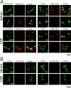
Fig 3. IgA antibodies to synapsin in serum and CSF of the previously described index patient with limbic encephalitis are directed against the D-domain of synapsin.
(A)HEK293 cells were transfected with eYFP-tagged synapsin I fragment ABC, eGFP-tagged DE, D or E. Cells were incubated with patient serum or CSF at a dilution of 1:320. Bound patient IgA was detected using a Texas red-coupled secondary antibody directed against human IgA. Both the patient’s serum and CSF (Patient IgA) demonstrated positive immunoreactivity that co-localized with the eGFP tag of synapsin DE and single D fragments, but with none of the other constructs. (B) Serum and CSF of two healthy controls (control IgA) did not bind to the cells transfected in the same way.
Fig 4
Fig 4. IgG subclasses of antibodies to synapsin in sera of patients with a clinically isolated syndrome (CIS), bipolar disorder and depression.
(A) HEK293 cells were transfected with eGFP-tagged full length rat synapsin Ia and incubated with serum of patient CIS117 at a dilution of 1:320. Bound antibodies of the IgG subclasses IgG1-4 were detected using subclass-specific Alexa 647-coupled secondary antibodies. The patient’s serum demonstrated positive immunoreactivity for IgG1 antibodies but none of the other subclasses. (B) HEK293 cells were transfected with eGFP-tagged full length rat synapsin Ia and incubated with serum of patient PSY 149 (upper two panels) and patient PSY 171 at a dilution of 1:320. Bound antibodies of the IgG subclasses IgG1-4 were detected as given in (A). Serum of patient PSY 149 demonstrated positive immunoreactivity for IgG1 and IgG4 antibodies but none of the other subclasses. Serum of patient PSY 171 demonstrated positive immunoreactivity for IgG3 antibodies but none of the other subclasses. (C) Serum of a healthy control did not exhibit immunoreactivity for any of the IgG subclasses.

References

    1. Piepgras J., Höltje M., Otto C., Harms H., Satapathy A., Cesca F., et al. Intrathecal immunoglobulin A and G antibodies to synapsin in a patient with limbic encephalitis. Neurol Neuroimmunol Neuroinflamm. 2015. November 4;2(6):e169 10.1212/NXI.0000000000000169 - DOI - PMC - PubMed
    1. Höltje M., Mertens R., Kochova E., Brix Schou M., Saether S.G., Prüss H., et al. Synapsin-antibodies in psychiatric and neurological disorders: prevalence and clinical finding. Brain, Behaviour and Immunity 2017; July 18 pii: S0889-1591(17)30218-0. 10.1016/j.immuni.2017.05.003 - DOI - PubMed
    1. Fassio A., Patry L., Congia S., Onofri F., Piton A., Gauthier J., et al. Syn1 loss-of-function mutations in autism and partial epilepsy cause impaired synaptic function. Hum Mol Genet 2011; 20:2297–307. 10.1093/hmg/ddr122 - DOI - PubMed
    1. Baldelli P., Fassio A., Valtorta F., Benfenati F. Lack of synapsin I reduces the readily releasable pool of synaptic vesicles at central inhibitory synapses. Journal of Neuroscience 2007; 27:13520–13531. 10.1523/JNEUROSCI.3151-07.2007 - DOI - PMC - PubMed
    1. Bragina L., Candiracci C., Barbaresi P., Giovedì S., Benfenati F., Conti F. Heterogeneity of glutamatergic and GABAergic release machinery in cerebral cortex. Neuroscience 2007; 146:1829–1840. 10.1016/j.neuroscience.2007.02.060 - DOI - PubMed

Publication types

MeSH terms