

Mechanisms of decreased cholinergic arousal in focal seizures: In vivo whole-cell recordings from the pedunculopontine tegmental nucleus
- PMID: 30543800
- PMCID: PMC6503533
- DOI: 10.1016/j.expneurol.2018.11.008
Mechanisms of decreased cholinergic arousal in focal seizures: In vivo whole-cell recordings from the pedunculopontine tegmental nucleus
Abstract
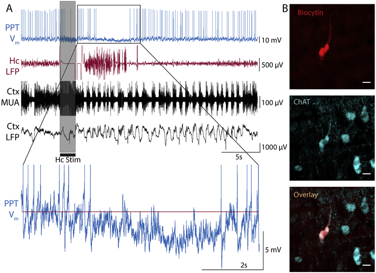
Focal limbic seizures often impair consciousness/awareness with major negative impact on quality of life. Recent work has shown that limbic seizures depress brainstem arousal systems, including reduced action potential firing in a key node: cholinergic neurons of the pedunculopontine tegmental nucleus (PPT). In vivo whole-cell recordings have not previously been achieved in PPT, but are used here with the goal of elucidating the mechanisms of reduced PPT cholinergic neuronal activity. An established model of focal limbic seizures was used in rats following brief hippocampal stimulation under light anesthesia. Whole-cell in vivo recordings were obtained from PPT neurons using custom-fabricated 9-10 mm tapered patch pipettes, and cholinergic neurons were identified histologically. Average membrane potential, input resistance, membrane potential fluctuations and variance were analyzed during seizures. A subset of PPT neurons exhibited reduced firing and hyperpolarization during seizures and stained positive for choline acetyltransferase. These PPT neurons showed a mean membrane potential hyperpolarization of -3.82 mV (±0.81 SEM, P < .05) during seizures, and also showed significantly increased input resistance, fewer excitatory post-synaptic potential (EPSP)-like events (P < .05), and reduced membrane potential variance (P < .01). The combination of increased input resistance, decreased EPSP-like events and decreased variance weigh against active ictal inhibition and support withdrawal of excitatory input as the dominant mechanism of decreased activity of cholinergic neurons in the PPT. Further identifying synaptic mechanisms of depressed arousal during seizures may lead to new treatments to improve ictal and postictal cognition.
Copyright © 2018 Elsevier Inc. All rights reserved.
Conflict of interest statement
Disclosure
None of the authors have any conflicts of interest to disclose. We confirm that we have read the Journal’s position on issues involved in ethical publication and affirm that this report is consistent with those guidelines.
Figures
Similar articles
-
The amygdala and the pedunculopontine tegmental nucleus: interactions controlling active (rapid eye movement) sleep.Exp Neurol. 2012 Nov;238(1):44-51. doi: 10.1016/j.expneurol.2012.08.001. Epub 2012 Aug 17. Exp Neurol. 2012. PMID: 22971360
-
Optogenetic stimulation of cholinergic brainstem neurons during focal limbic seizures: Effects on cortical physiology.Epilepsia. 2015 Dec;56(12):e198-202. doi: 10.1111/epi.13220. Epub 2015 Nov 4. Epilepsia. 2015. PMID: 26530287 Free PMC article.
-
Noradrenaline excites non-cholinergic laterodorsal tegmental neurons via two distinct mechanisms.Neuroscience. 1999;93(2):619-30. doi: 10.1016/s0306-4522(99)00130-x. Neuroscience. 1999. PMID: 10465446
-
Decreased subcortical cholinergic arousal in focal seizures.Neuron. 2015 Feb 4;85(3):561-72. doi: 10.1016/j.neuron.2014.12.058. Neuron. 2015. PMID: 25654258 Free PMC article.
-
Alpha-2 adrenergic regulation of pedunculopontine nucleus neurons during development.Neuroscience. 2006 Aug 25;141(2):769-779. doi: 10.1016/j.neuroscience.2006.04.045. Epub 2006 Jun 6. Neuroscience. 2006. PMID: 16753270
Cited by
-
A review of cell-type specific circuit mechanisms underlying epilepsy.Acta Epileptol. 2024 Jun 1;6(1):18. doi: 10.1186/s42494-024-00159-2. Acta Epileptol. 2024. PMID: 40217549 Free PMC article. Review.
-
Comparison of anaesthetic- and seizure-induced states of unconsciousness: a narrative review.Br J Anaesth. 2021 Jan;126(1):219-229. doi: 10.1016/j.bja.2020.07.056. Epub 2020 Sep 18. Br J Anaesth. 2021. PMID: 32951841 Free PMC article. Review.
-
Huperzine A attenuates epileptic seizures via enhancing dCA1-projecting septal cholinergic transmission.Acta Pharmacol Sin. 2025 Aug;46(8):2151-2162. doi: 10.1038/s41401-025-01522-w. Epub 2025 Mar 26. Acta Pharmacol Sin. 2025. PMID: 40140526
-
Up and Down States of Cortical Neurons in Focal Limbic Seizures.Cereb Cortex. 2020 May 14;30(5):3074-3086. doi: 10.1093/cercor/bhz295. Cereb Cortex. 2020. PMID: 31800015 Free PMC article.
-
A Century Searching for the Neurons Necessary for Wakefulness.Front Neurosci. 2022 Jul 19;16:930514. doi: 10.3389/fnins.2022.930514. eCollection 2022. Front Neurosci. 2022. PMID: 35928009 Free PMC article. Review.
References
-
- Benarroch EE, 2013. Pedunculopontine nucleus: functional organization and clinical implications. Neurology 80, 1148–1155. - PubMed
-
- Blumenfeld H, McNally KA, Vanderhill SD, Paige AL, Chung R, Davis K, Norden AD, Stokking R, Studholme C, Novotny EJ, Zubal IG, Spencer SS, 2004. Positive and negative network correlations in temporal lobe epilepsy. Cerebral Cortex 14, 892–902. - PubMed
-
- Chance FS, Abbott L, Reyes AD, 2002. Gain modulation from background synaptic input. Neuron 35, 773–782. - PubMed
-
- Destexhe A, Paré D, 1999. Impact of network activity on the integrative properties of neocortical pyramidal neurons in vivo. Journal of neurophysiology 81, 1531–1547. - PubMed
Publication types
MeSH terms
Substances
Grants and funding
LinkOut - more resources
Full Text Sources
Medical
