

doi: 10.1016/j.jcmgh.2018.11.001.
Epub 2018 Dec 10.
Human Norovirus Propagation in Human Induced Pluripotent Stem Cell-Derived Intestinal Epithelial Cells
Affiliations
- PMID: 30543870
- PMCID: PMC6477164
- DOI: 10.1016/j.jcmgh.2018.11.001
Item in Clipboard
Human Norovirus Propagation in Human Induced Pluripotent Stem Cell-Derived Intestinal Epithelial Cells
Cell Mol Gastroenterol Hepatol.
2019.
No abstract available
Figures
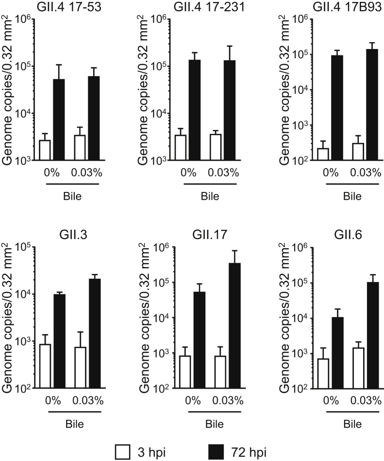
Replication of HuNoVs in IECs derived from human iPSCs. Monolayered human iPSC–derived IECs were inoculated with 2 × 10⁶ genome equivalents of the indicated HuNoV genotypes. Inoculation and sampling were done as described in the Materials and Methods. Viral genome RNA was extracted from both supernatants (ie, those taken at 3 and 72 hours postinfection [hpi]), and then genome equivalents were quantified with reverse transcriptase quantitative polymerase chain reaction. Samples at 3 hpi were used as references. Each value is representative of at least 3 independent experiments and is shown as the mean ± SD from between 4 and 6 wells of supernatants of each culture group.
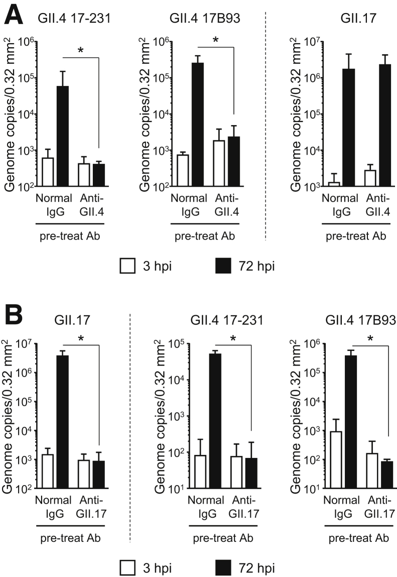
Replication of GII.4 genotype HuNoVs is prevented by pretreatment with either anti-GII.4 or anti-GII.17 pAbs. Before inoculation of IECs, 2 × 10⁶ genome equivalents of each HuNoV genotype was incubated with 100 ng of (A) anti-GII.4 or (B) anti-GII.17 Ab, or with normal rabbit immunoglobulin G, for 1.5 hours. Inoculation, sampling, and quantification of genome equivalents were done as described in the Materials and Methods. Each value is representative of at least 3 independent experiments and is shown as the mean ± SD from between 4 and 6 wells of supernatants of each culture group. *P < .05. hpi, hours postinfection.
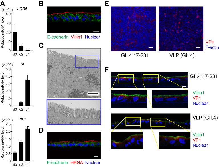
Polarized monolayer of IECs derived from human iPSCs. (A) Differentiation of iPSC-derived IECs to enterocytes. Relative messenger RNA expression of the indicated genes in iPSC-derived IECs during the course of differentiation (days 0, 2, and 4) were determined by qPCR and normalized against the expression of GAPDH. The assays were performed in 3 independent biological replicates. Expression levels are presented as means ± SEM. SI, sucrase isomaltase. (B, D) Immunohistochemical analysis of monolayers harvested with the Transwell membrane after 6 days of culture. Sections were stained with anti-E-cadherin (green) and anti-villin-1 antibodies (red, B) or Ulex europaeus agglutinin-1 (red, D), and counterstained with DAPI (blue). Data are representative of 3 independent experiments. Scale bar = 10 μm. HBGA, histo-blood group antigen. (C) Transmission electron microscopic images of polarized monolayer of human iPSC–derived IECs. Lower panel is a magnification of the area in the blue box in the upper panel. Data are representative of 3 independent experiments. Scale bar = 2.5 μm. (E, F) GII.4 virus and VLPs bound to, and entered, the iPSC-derived IECs. Polarized iPSC-derived IECs on Transwell membranes were incubated with 8.3 × 10⁸ genome equivalents of GII.4 virus or 300 ng of VLPs for 3 hours, and then the Transwell membranes were whole-mount stained with anti-GII.4 antibody (E, red) or were sectioned and stained with anti-GII.4 antibody (F, red) simultaneously with anti-villin-1 antibody (F, green), and then counterstained with DAPI (blue). Lower panels are a magnification of the area in the yellow boxes in the upper panels. Data are representative of three independent experiments. Scale bar = 50 μm (E), 10 μm (F).
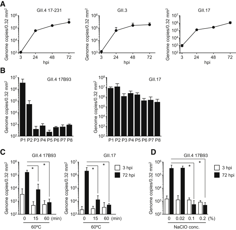
Inactivation of HuNoVs by heat and sodium hypochlorite treatment. Monolayered human iPSC–derived IECs were inoculated with (A) 2 × 10⁶ or (B) 3.7 × 10⁷ (GII.4) or 5.8 × 10⁸ (GII.17) genome equivalents of the indicated HuNoV genotypes. Inoculation and sampling were done as described in the Materials and Methods. Viral genome RNA was extracted from each supernatant sampled at (A) the indicated time or (B) 24 to 48 hpi, and then genome equivalents were quantified with reverse transcriptase qPCR. Each value is representative of at least 3 independent experiments and is shown as the mean ± SD from (A) 6 or (B) 3 wells of supernatants of each culture group. (C) GII.4 and GII.17 HuNoVs (2 × 10⁶ genome equivalents) were incubated at 60°C for the indicated times. (D) GII.4 HuNoVs (2 × 10⁷ genome equivalents) were incubated with the indicated concentrations of sodium hypochlorite (NaClO) at room temperature for 30 minutes, and then diluted with base medium to 2 × 10⁶ genome equivalents/100 μL. Monolayered human iPSC–derived IECs were inoculated with each treated HuNoV. Viral genome RNA was extracted from both supernatants, and the genome equivalents were quantified by reverse transcriptase qPCR. Samples at 3 hpi were used as in the references (A, C, and D). Each value is representative of 3 independent experiments and is shown as the mean ± SD from 6 wells of supernatants of each culture group. *P < .05.
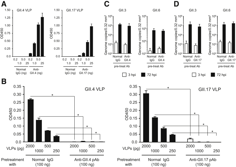
Binding of GII.4 and GII.17 VLPs to histo-blood group antigens is prevented by pre-treatment with each anti-VLP polyclonal antibody (pAb). (A) Homotypic titer of anti-GII.4 and GII.17 pAbs were quantified by enzyme-linked immnosorbent assay. Data are means ± SD from 1 experiment representative of 2 independent experiments. (B) Blocking activity of anti-GII.4 and anti-GII.17 toward VLP–histo-blood group antigen binding was measured by ELISA. Data are mean ± SD from 1 experiment representative of 3 independent experiments. *P < .05. GII.3 and GII.6 HuNoVs (2 × 10⁶ genome equivalents) were incubated with 100 ng of (C) anti-GII.4 or (D) anti-GII.17 antibody, or with normal rabbit IgG, for 1.5 hours. Monolayered human iPSC–derived IECs were inoculated with each treated HuNoV. Viral genome RNA was extracted from both supernatants, and the genome equivalents were quantified by reverse transcriptase qPCR. Samples at 3 hpi were used as references. Each value is representative of 3 independent experiments and is shown as the mean ± SD from 4 to 6 wells of supernatants of each culture group.
Similar articles
-
Alcohol abrogates human norovirus infectivity in a pH-dependent manner.Sci Rep. 2020 Sep 28;10(1):15878. doi: 10.1038/s41598-020-72609-z. Sci Rep. 2020. PMID: 32985508 Free PMC article.
-
Simultaneous comparison of murine norovirus, feline calicivirus, coliphage MS2, and GII.4 norovirus to evaluate the efficacy of sodium hypochlorite against human norovirus on a fecally soiled stainless steel surface.Foodborne Pathog Dis. 2011 Sep;8(9):1005-10. doi: 10.1089/fpd.2010.0782. Epub 2011 Apr 2. Foodborne Pathog Dis. 2011. PMID: 21457050
-
Human Norovirus Replication in Human Intestinal Enteroids as Model to Evaluate Virus Inactivation.Emerg Infect Dis. 2018 Aug;24(8):1453-1464. doi: 10.3201/eid2408.180126. Emerg Infect Dis. 2018. PMID: 30014841 Free PMC article.
-
Human Norovirus Cultivation in Nontransformed Stem Cell-Derived Human Intestinal Enteroid Cultures: Success and Challenges.Viruses. 2019 Jul 11;11(7):638. doi: 10.3390/v11070638. Viruses. 2019. PMID: 31336765 Free PMC article. Review.
-
[Current topics on inactivation of norovirus].Kokuritsu Iyakuhin Shokuhin Eisei Kenkyusho Hokoku. 2011;(129):37-54. Kokuritsu Iyakuhin Shokuhin Eisei Kenkyusho Hokoku. 2011. PMID: 22259842 Review. Japanese.
Cited by
-
Lactobacilli as a Vector for Delivery of Nanobodies against Norovirus Infection.Pharmaceutics. 2022 Dec 25;15(1):63. doi: 10.3390/pharmaceutics15010063. Pharmaceutics. 2022. PMID: 36678692 Free PMC article.
-
Simultaneous Immunization with Multivalent Norovirus VLPs Induces Better Protective Immune Responses to Norovirus Than Sequential Immunization.Viruses. 2019 Nov 2;11(11):1018. doi: 10.3390/v11111018. Viruses. 2019. PMID: 31684058 Free PMC article.
-
Advances in Human Norovirus Vaccine Research.Vaccines (Basel). 2021 Jul 2;9(7):732. doi: 10.3390/vaccines9070732. Vaccines (Basel). 2021. PMID: 34358148 Free PMC article. Review.
-
Norovirus replication, host interactions and vaccine advances.Nat Rev Microbiol. 2025 Jun;23(6):385-401. doi: 10.1038/s41579-024-01144-9. Epub 2025 Jan 17. Nat Rev Microbiol. 2025. PMID: 39824927 Free PMC article. Review.
-
Current trends and new approaches for human norovirus replication in cell culture: a literature review.Arch Virol. 2024 Mar 8;169(3):71. doi: 10.1007/s00705-024-05999-4. Arch Virol. 2024. PMID: 38459228 Review.
References
Publication types
MeSH terms
Substances
LinkOut - more resources
Full Text Sources
Other Literature Sources
Medical
