. 2019 Feb 20;47(3):1082-1096.
doi: 10.1093/nar/gky1239.

Diverse lipid conjugates for functional extra-hepatic siRNA delivery in vivo

Affiliations

Diverse lipid conjugates for functional extra-hepatic siRNA delivery in vivo

Annabelle Biscans et al. Nucleic Acids Res. .

Abstract

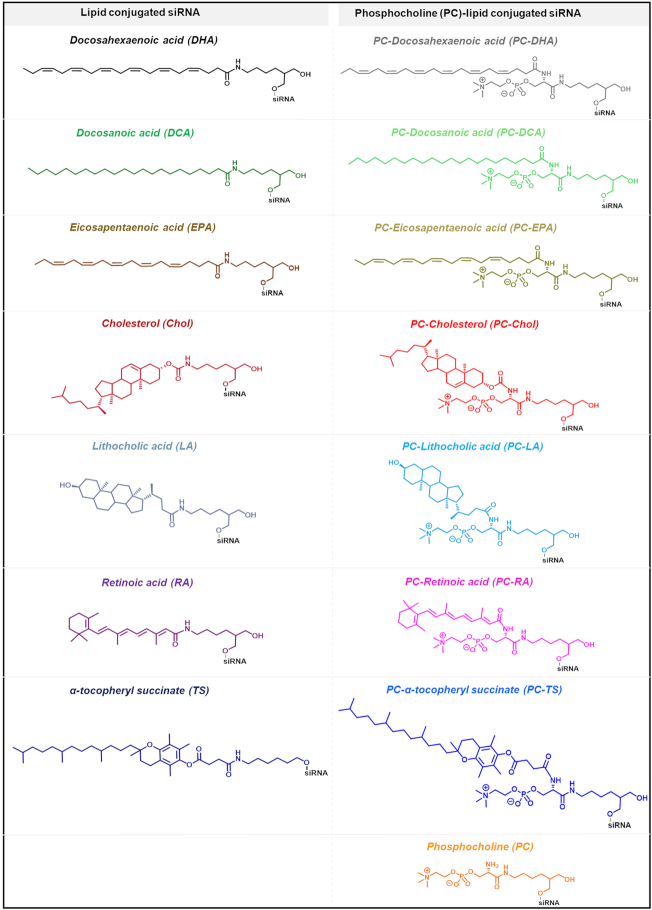
Small interfering RNA (siRNA)-based therapies are proving to be efficient for treating liver-associated disorders. However, extra-hepatic delivery remains challenging, limiting therapeutic siRNA utility. We synthesized a panel of fifteen lipid-conjugated siRNAs and systematically evaluated the impact of conjugate on siRNA tissue distribution and efficacy. Generally, conjugate hydrophobicity defines the degree of clearance and the liver-to-kidney distribution profile. In addition to primary clearance tissues, several conjugates achieve significant siRNA accumulation in muscle, lung, heart, adrenal glands and fat. Oligonucleotide distribution to extra-hepatic tissues with some conjugates was significantly higher than with cholesterol, a well studied conjugate, suggesting that altering conjugate structure can enhance extra-hepatic delivery. These conjugated siRNAs enable functional gene silencing in lung, muscle, fat, heart and adrenal gland. Required levels for productive silencing vary (5-200 μg/g) per tissue, suggesting that the chemical nature of conjugates impacts tissue-dependent cellular/intracellular trafficking mechanisms. The collection of conjugated siRNA described here enables functional gene modulation in vivo in several extra-hepatic tissues opening these tissues for gene expression modulation. A systemic evaluation of a panel of conjugated siRNA, as reported here, has not previously been investigated and shows that chemical engineering of lipid siRNAs is essential to advance the RNA therapeutic field.


Figures

Figure 1.
Figure 1.
Library of studied lipid conjugated siRNAs. The desired conjugate is attached to the 3′ end of the sense strand of the siRNA though a C7 linker (left column) or a C7 linker functionalized with a phosphocholine group (PC) (right column).
Figure 2.
Figure 2.
Experimental plan describing the study of one conjugated siRNA in mice. The experiment was performed for 15 conjugated and one unconjugated siRNA. In total, 321 mice were injected (included PBS-injected controls). For distribution studies, three mice per lipid-conjugated siRNAHtt were sacrificed. Fifteen tissues per mouse (liver, kidneys, lung, heart, thymus, spleen, pancreas, adrenal glands, intestine, fallopian tube, bladder, fat, muscle, skin at injection site and skin away from injection site) were collected and analyzed by fluorescence microscopy and High Performance Liquid Chromatography for antisense strand quantification using a PNA hybridization assay. For efficacy studies, eight mice injected with conjugated siRNAHtt and eight mice injected with conjugated siRNAPpibper lipid conjugate were sacrificed. The expression of Htt mRNA or Ppib mRNA in tissues were measured using QuantiGene Assay.
Figure 3.
Figure 3.
The presence of fluorophore Cy3 on the lipid-conjugated siRNA does not significantly affect siRNA distribution in vivo. Tissue accumulation levels of (A) Cy3-labeled and unlabeled DCA-conjugated siRNA (B) or Cy3-labeled and unlabeled PC-DCA conjugated siRNA were similar in 15 tissues. siRNA quantification was performed by PNA hybridization assay (average of three mice per conjugate ± SD) after a single subcutaneous injection of 20 mg/kg conjugated siRNA and tissue collection after 48 h.
Figure 4.
Figure 4.
The conjugate defines the ratio of liver to kidney distribution. (A) Representative fluorescence images of kidney and liver sections from mice injected subcutaneously with 20 mg/kg Cy3-labeled lipid-conjugated siRNAs. Nuclei stained with DAPI. Three mice per conjugate were injected and tissue collected 48 h after injection. Images taken at 5 × magnification and collected at the same laser intensity and acquisition time. Scale, 1 mm. (B) siRNA quantification in kidney (cortex) and liver was performed by PNA hybridization assay (average of three mice per conjugate ± SD).
Figure 5.
Figure 5.
The conjugate impacts the cell-type distribution in kidney and liver. Representative fluorescence images of (A) kidney cortex and kidney medulla or (B) liver sections from mice injected subcutaneously with 20 mg/kg Cy3-labeled lipid-conjugated siRNAs. Nuclei stained with DAPI. Three mice per conjugate were injected and tissue collected 48 h after injection. Images taken at 40 × magnification and collected at the same laser intensity and acquisition time. Scale, 50 μm.
Figure 6.
Figure 6.
The nature of the lipid conjugate has a profound impact on siRNA tissue retention. Bar graph showing percent of the injected dose cumulatively retained in mice across all 15 tissues 48 h after a single subcutaneous injection of 20 mg/kg conjugated siRNA (average of n = 3 mice). Stacked bars indicate the percent of the retained dose in liver, kidney, site of injection and others tissues. More than 80% of unconjugated siRNAs are cleared while highly hydrophobic compounds are retained quantitatively.
Figure 7.
Figure 7.
Conjugated siRNAs accumulate into extra-hepatic tissues, accumulation defines by the structure of the conjugate. Bar graph showing the quantity of the antisense strand of conjugated siRNAs present in heart, lung, muscle and fat 48 h after a single subcutaneous injection with 20 mg/kg (n = 3 mice per conjugate ± SD). siRNA quantification was measured by PNA hybridization assay. DCA and PC-DCA conjugates improve extra-hepatic siRNA accumulation compared to cholesterol.
Figure 8.
Figure 8.
Relative improvement of DCA conjugated siRNA extra-hepatic tissue accumulation compared to cholesterol conjugated siRNA. Bar graph showing the ratio between the quantity of the DCA-siRNA antisense strands and cholesterol-siRNA antisense strands present in 15 tissues 48 h after a single subcutaneous injection with 20 mg/kg (n = 3 mice per conjugate ± SD). siRNA quantification was measured by PNA hybridization assay. DCA accumulation was 3- to 4-fold higher in heart, muscle and fat, ∼6-fold higher in skin (systemic) and 9-fold higher in lung.
Figure 9.
Figure 9.
Accumulation of conjugated siRNA in several extra-hepatic tissues such as heart, muscle, fat, adrenal glands and lung are sufficient to induce mRNA silencing. Representative fluorescence images of heart, muscle, fat, adrenal glands and lung sections from mice injected subcutaneously with 20 mg/kg Cy3-labeled lipid-conjugated siRNAs or PBS. Nuclei stained with DAPI. Three mice per conjugate were injected and tissue collected after 48 h. Images taken at 40 × magnification and collected at the same laser intensity and acquisition time. Scale, 50 μm. For the measurement of Huntingtin (Htt) or Cyclophilin B (Ppib) mRNA levels, mice were injected subcutaneously with 20 mg/kg of conjugated siRNA (n = 16 per conjugate: 8 with siRNA targeting Htt and 8 with siRNA targeting Ppib, as well as non-targeting controls). The tissues were collected after 1 week and mRNA levels were measured using QuantiGene® (Affymetrix), normalized to a housekeeping gene, Hprt (Hypoxanthine-guanine phosphoribosyl transferase) and presented as percent of PBS control (mean ± SD).
Figure 10.
Figure 10.
The efficiency of target mRNA silencing does not necessarily correlate with siRNA accumulation level. Graphs showing target mRNA silencing (relative to PBS) for different conjugates with similar tissue accumulation (right Y-axis, bar). Mice were injected subcutaneously with 20 mg/kg lipid-conjugated siRNAs (n = 3 ± SD, 48 h for antisense strand quantification; n = 8 ± SD, 1 week for target gene silencing QuantiGene). While several conjugates show similar tissue accumulation, the functional silencing is conjugate-specific.

References

    1. Zhou J., Shum K.-T., Burnett J.C., Rossi J.J.. Nanoparticle-based delivery of RNAi therapeutics: progress and challenges. Pharmaceuticals. 2013; 6:85–107. - PMC - PubMed
    1. Nair J.K., Willoughby J.L.S., Chan A., Charisse K., Alam M.R., Wang Q., Hoekstra M., Kandasamy P., Kel’in A.V., Milstein S. et al. . Multivalent N-Acetylgalactosamine-Conjugated siRNA localizes in hepatocytes and elicits robust RNAi-mediated gene silencing. JACS. 2014; 136:16958–16961. - PubMed
    1. Zimmermann T.S., Karsten V., Chan A., Chiesa J., Boyce M., Bettencourt B.B., Hutabarat R., Nochur S., Vaishnaw A., Gollob J.. Clinical proof of concept for a novel hepatocyte-targeting GalNAc-siRNA conjugate. Mol. Ther. 2017; 25:71–78. - PMC - PubMed
    1. Hassler M.R., Turanov A.A., Alterman J.F., Haraszti R.A., Coles A.H., Osborn M.F., Echeverria D., Nikan M., Salomon W.E., Roux L. et al. . Comparison of partially and fully chemically-modified siRNA in conjugate-mediated delivery in vivo. Nucleic Acids Res. 2018; 46:2185–2196. - PMC - PubMed
    1. Nair J.K., Attarwala H., Sehgal A., Wang Q., Aluri K., Zhang X., Gao M., Liu J., Indrakanti R., Schofield S. et al. . Impact of enhanced metabolic stability on pharmacokinetics and pharmacodynamics of GalNAc–siRNA conjugates. Nucleic Acids Res. 2017; 45:10969–10977. - PMC - PubMed

Publication types