

Metanephric adenoma with low apparent diffusion coefficient value mimicking renal cell carcinoma: A case report
- PMID: 30544462
- PMCID: PMC6310553
- DOI: 10.1097/MD.0000000000013539
Metanephric adenoma with low apparent diffusion coefficient value mimicking renal cell carcinoma: A case report
Abstract
Rationale: Metanephric adenoma (MA) is a rare and often benign tumor. Most MAs were misdiagnosed as renal cell carcinomas (RCCs) preoperatively. Diffusion weighted imaging (DWI) and apparent diffusion coefficient (ADC) mapping can help to differentiate benign and malignant tumors. However, there are still pitfalls in using DWI and ADC to discriminate benign and malignant lesions.
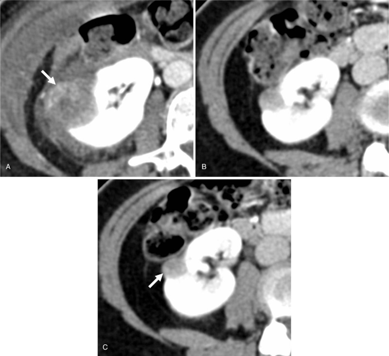
Patient concerns: A 56-year-old woman had a right renal metanephric adenoma. The tumor showed very low ADC value preoperatively and was misdiagnosed as a renal cell carcinoma.
Diagnosis: Intraoperative ultrasound-guided percutaneous biopsy of tumor was performed. Based on the histopathological findings and immunohistochemical stains, a diagnosis of metanephric adenoma was suggested.
Interventions: The patient received percutaneous cryoablation of this tumor. Five years later, she underwent right partial nephrectomy because local recurrence was revealed on a follow-up computed tomography (CT).
Outcomes: MA was confirmed again by histological examination. The patient was uneventful after surgery.
Lessons: ADC mapping can be used for differentiating RCCs from other benign tumors by their lower ADC values. However, some benign and malignant lesions have overlapped low ADC values. This case illustrated that a benign lesion such as MA could mimic RCC on ADC, by its highly cellular component. Cryoablation is an optional treatment, which has an increased risk of local recurrence. Follow-up CT or MRI is useful and necessary for detection of local recurrence by depicting enhancing solid parts in a tumor over time.
Conflict of interest statement
The authors report no conflicts of interest
Figures


Similar articles
-
Differential diagnosis of the small renal masses: role of the apparent diffusion coefficient of the diffusion-weighted MRI.Int Urol Nephrol. 2018 Feb;50(2):197-204. doi: 10.1007/s11255-017-1761-1. Epub 2017 Dec 11. Int Urol Nephrol. 2018. PMID: 29230706
-
Renal metanephric adenoma mimicking papillary renal cell carcinoma on computed tomography: a case report.Urol Int. 2013;90(3):369-72. doi: 10.1159/000341940. Epub 2012 Oct 17. Urol Int. 2013. PMID: 23076029
-
Subtype Differentiation of Small (≤ 4 cm) Solid Renal Mass Using Volumetric Histogram Analysis of DWI at 3-T MRI.AJR Am J Roentgenol. 2018 Sep;211(3):614-623. doi: 10.2214/AJR.17.19278. Epub 2018 May 29. AJR Am J Roentgenol. 2018. PMID: 29812980
-
Metanephric adenoma in an 8-year-old child: case report and review of the literature.J Pediatr Surg. 2005 May;40(5):e25-8. doi: 10.1016/j.jpedsurg.2005.02.019. J Pediatr Surg. 2005. PMID: 15937802 Review.
-
Metanephric Adenoma in the Pediatric Population: Diagnostic Challenges and Follow-up.Urology. 2018 Oct;120:211-215. doi: 10.1016/j.urology.2018.06.042. Epub 2018 Jul 10. Urology. 2018. PMID: 30006267 Review.
References
-
- Amin MB, Amin MB, Tamboli P, et al. Prognostic impact of histologic subtyping of adult renal epithelial neoplasms: an experience of 405 cases. Am J Surg Pathol 2002;26:281–91. - PubMed
-
- Davis CJ, Barton JH, Sesterhenn IA, et al. Metanephric adenoma: clinicopathological study of fifty patients. Am J Surg Pathol 1995;19:1101–14. - PubMed
-
- Lassel EA, Rao R, Schwenke C, et al. Diffusion-weighted imaging of focal renal lesions: a meta-analysis. Eur Radiol 2014;24:241–9. - PubMed
-
- Eble JN, Sauter G, Epstein JI, et al. Pathology and Genetics. Tumors of the Urinary System and Male Genital Organs. Lyon: IARC Press; 2004.
-
- Hartman DJ, Maclennan GT. Renal metanephric adenoma. J Urol 2007;178(3 pt 1):1058. - PubMed
Publication types
MeSH terms
LinkOut - more resources
Full Text Sources
Medical
