

Tendon Remodeling in Response to Resistance Training, Anabolic Androgenic Steroids and Aging
- PMID: 30544536
- PMCID: PMC6316563
- DOI: 10.3390/cells7120251
Tendon Remodeling in Response to Resistance Training, Anabolic Androgenic Steroids and Aging
Abstract
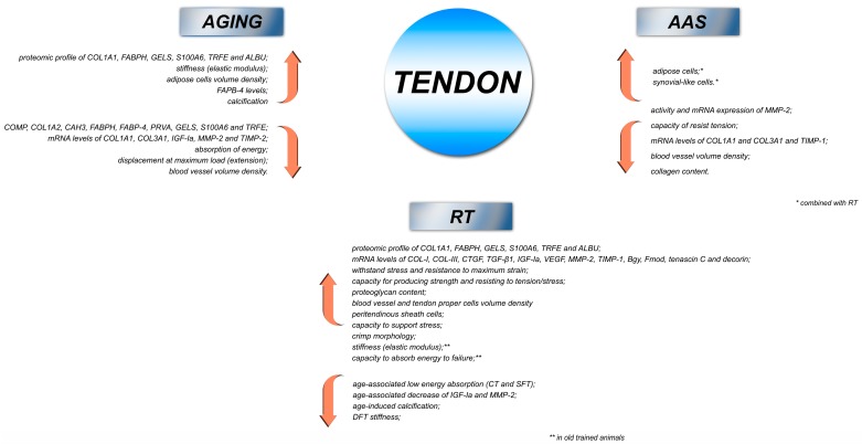
Exercise training (ET), anabolic androgenic steroids (AAS), and aging are potential factors that affect tendon homeostasis, particularly extracellular matrix (ECM) remodeling. The goal of this review is to aggregate findings regarding the effects of resistance training (RT), AAS, and aging on tendon homeostasis. Data were gathered from our studies regarding the impact of RT, AAS, and aging on the calcaneal tendon (CT) of rats. We demonstrated a series of detrimental effects of AAS and aging on functional and biomechanical parameters, including the volume density of blood vessel cells, adipose tissue cells, tendon calcification, collagen content, the regulation of the major proteins related to the metabolic/development processes of tendons, and ECM remodeling. Conversely, RT seems to mitigate age-related tendon dysfunction. Our results suggest that AAS combined with high-intensity RT exert harmful effects on ECM remodeling, and also instigate molecular and biomechanical adaptations in the CT. Moreover, we provide further information regarding the harmful effects of AAS on tendons at a transcriptional level, and demonstrate the beneficial effects of RT against the age-induced tendon adaptations of rats. Our studies might contribute in terms of clinical approaches in favor of the benefits of ET against tendinopathy conditions, and provide a warning on the harmful effects of the misuse of AAS on tendon development.
Keywords: aging; anabolic androgenic steroids; extracellular matrix; resistance training; tendon.
Conflict of interest statement
The authors declare no conflict of interest.
Figures
Similar articles
-
Androgenic-anabolic steroids associated with mechanical loading inhibit matrix metallopeptidase activity and affect the remodeling of the achilles tendon in rats.Am J Sports Med. 2006 Aug;34(8):1274-80. doi: 10.1177/0363546506286867. Epub 2006 Apr 24. Am J Sports Med. 2006. PMID: 16636352
-
Detraining and Anabolic-Androgenic Steroid Discontinuation Change Calcaneal Tendon Morphology.J Funct Morphol Kinesiol. 2018 Dec 21;4(1):1. doi: 10.3390/jfmk4010001. J Funct Morphol Kinesiol. 2018. PMID: 33467316 Free PMC article.
-
Tendon structural adaptations to load exercise are inhibited by anabolic androgenic steroids.Scand J Med Sci Sports. 2014 Feb;24(1):e39-51. doi: 10.1111/sms.12135. Epub 2013 Nov 14. Scand J Med Sci Sports. 2014. PMID: 24224869
-
Anabolic steroids and tendons: A review of their mechanical, structural, and biologic effects.J Orthop Res. 2018 Nov;36(11):2830-2841. doi: 10.1002/jor.24116. Epub 2018 Sep 5. J Orthop Res. 2018. PMID: 30047601
-
Effects of androgenic-anabolic steroids in athletes.Sports Med. 2004;34(8):513-54. doi: 10.2165/00007256-200434080-00003. Sports Med. 2004. PMID: 15248788 Review.
Cited by
-
Effects of aging on the histology and biochemistry of rat tendon healing.BMC Musculoskelet Disord. 2021 Nov 15;22(1):949. doi: 10.1186/s12891-021-04838-w. BMC Musculoskelet Disord. 2021. PMID: 34781961 Free PMC article.
-
Application of Orthobiologics in Achilles Tendinopathy: A Review.Life (Basel). 2022 Mar 9;12(3):399. doi: 10.3390/life12030399. Life (Basel). 2022. PMID: 35330150 Free PMC article. Review.
-
Intra-Articular Hyaluronic Acid in Osteoarthritis and Tendinopathies: Molecular and Clinical Approaches.Biomedicines. 2023 Mar 30;11(4):1061. doi: 10.3390/biomedicines11041061. Biomedicines. 2023. PMID: 37189679 Free PMC article. Review.
-
Anabolic Androgenic Steroids in Orthopaedic Surgery: Current Concepts and Clinical Applications.J Am Acad Orthop Surg Glob Res Rev. 2022 Jan 4;6(1):e21.00156. doi: 10.5435/JAAOSGlobal-D-21-00156. J Am Acad Orthop Surg Glob Res Rev. 2022. PMID: 34982051 Free PMC article.
-
Effect of Aging on Tendon Biology, Biomechanics and Implications for Treatment Approaches.Int J Mol Sci. 2023 Oct 14;24(20):15183. doi: 10.3390/ijms242015183. Int J Mol Sci. 2023. PMID: 37894875 Free PMC article. Review.
References
-
- Kader D., Mosconi M., Benazzo F., Maffulli N. Tendon Injuries. Springer; London, UK: 2005. Achilles tendon rupture.
Publication types
Grants and funding
- 88887.136367/2017-00/Coordenação de Aperfeiçoamento de Pessoal de Nível Superior/International
- 465699/2014-6, 445069/2014-7/Conselho nacional de Pesquisa - CNPq/International
- 2013/00798-2, 2014/50938-8/Fundação de Amparo à Pesquisa do Estado de São Paulo/International
- 193.000.653/2015/Fundação de Apoio à Pesquisa do Distrito Federal/International
LinkOut - more resources
Full Text Sources
