. 2018 Dec 7;7(12):254.
doi: 10.3390/cells7120254.

Modulation of Protein Synthesis by eIF2α Phosphorylation Protects Cell from Heat Stress-Mediated Apoptosis

Affiliations

Modulation of Protein Synthesis by eIF2α Phosphorylation Protects Cell from Heat Stress-Mediated Apoptosis

Soyoung Park et al. Cells. .

Abstract

Global warming poses a considerable threat to human health, necessitating a proper understanding of mechanisms underlying cell death in the pathogenesis of heat-related diseases. Although mechanisms governing cytoplasmic response to heat are well understood, processes regulating cellular response to disruption of proteostasis in the endoplasmic reticulum (ER) due to heat stress remain unclear. The current study reveals that hyperthermic conditions may lead to a disturbance of ER homeostasis, also known as ER stress. Subsequent activation of the unfolded protein response (UPR) resulted in concomitant induction of cell death. Among the three UPR signaling pathways, the eIF2α phosphorylation pathway, and not the IRE1α/ATF6α pathways, is likely the main contributor to cell death under heat stress. Considering the role of eIF2α in translational control, we investigated the protective effect of translation rate on heat stress-mediated cell death. When protein synthesis was attenuated using cycloheximide or homoharringtonine, cell death due to heat stress was significantly reduced. In summation, we propose that transient modulation of protein synthesis by eIF2α phosphorylation has a pivotal role in protecting cells from heat stress-induced apoptosis. Therefore, pharmacological agents that promote eIF2α phosphorylation or reduce ER stress may contribute to the development of promising therapeutic approaches against heat-related diseases.

Keywords: ER stress; eIF2α phosphorylation; heat stress; translation; unfolded protein response (UPR).


Conflict of interest statement

All the authors declare no competing interests.

Figures
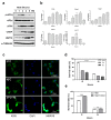
Figure 1
Figure 1
High temperature induces endoplasmic reticulum (ER) stress-mediated cell death. (a) Protein expression under condition of hyperthermia was measured. Cell lysates were collected at indicated times following incubation at 42 °C or tunicamycin (Tm) (1 μg/mL) treatment for western blot analysis. (b) Effect of heat stress on the unfolded protein response (UPR) gene expression. Quantitative RT-PCR was performed using total RNA extracted from mouse embryonic fibroblasts (MEFs) which were incubated at 42 °C for indicated time periods. Data are presented as means ± SEM (n = 3 independent experiments). (c) KDEL and DAPI immunofluorescence staining. KDEL and DAPI staining were performed using MEFs incubated at 42 °C or treated with thapsigargin (Tg) (300 nM) for 12 h. Representative images are shown. Scale bar represents 100 μm. (d) Cell viability was measured in MEFs treated with Tg (300 nM), Tm (1 μg/mL), or incubated at 42 °C for the indicated time periods. (e) Cell viability was measured in MEFs which were incubated at 42 °C for indicated time periods in the presence or absence of tauroursodeoxycholic acid (TUDCA) (2.4 mM). ** p < 0.01; **** p < 0.0001.
Figure 2
Figure 2
The IRE1α pathway does not protect cells from heat stress-mediated death. (a) Quantitative RT-PCR was performed using total RNA, extracted from Ire1α−/− and Ire1α+/+ MEFs which were incubated at 42 °C for indicated time periods. Data are presented as means ± SEM. (n = 3 independent experiments). (b) Protein expression under conditions of hyperthermia was measured. Following incubation at 42 °C, cell lysates were collected at the indicated times for western blot analysis. (c) Cell viability was measured in Ire1α−/− and Ire1α+/+ MEFs which were incubated at 42 °C for indicated time periods. (d) Caspase 3/7 activity was detected in Ire1α−/− and Ire1α+/+ MEFs which were incubated at 42 °C for 12 h. (e) Expression of sXBP1 (spliced form) and tXBP (total form) were measured using MEFs treated with 4μ8c at indicated doses in the presence or absence of Tg (300 nM) for 12 h. (f) Protein expression under conditions of hyperthermia was measured in the presence or absence of 4μ8c (20 μM). (g) Cell viability was measured in Ire1α−/− and Ire1α+/+ MEFs which were incubated at 42 °C for indicated time periods in the presence or absence of 4μ8c (20 μM). * p < 0.05; ** p < 0.01; *** p < 0.001; **** p < 0.0001.
Figure 3
Figure 3
The ATF6α pathway does not protect cells from heat stress-mediated death. (a) Quantitative RT-PCR was performed using total RNA extracted from Atf6α−/− and Atf6α+/+ MEFs which were incubated at 42 °C for indicated time periods. Data are presented as means ± SEM (n = 3 independent experiments). (b) Protein expression under conditions of hyperthermia was measured. Following 42 °C incubation, cell lysates were collected at indicated times for western blot analysis. (c) Cell viability was measured in Atf6α−/− and Atf6α+/+ MEFs following incubation at 42 °C for indicated time periods. (d) Caspase 3/7 activity was detected in Atf6α−/− and Atf6α+/+ MEFs which were incubated at 42 °C for 12 h. ** p < 0.01; *** p < 0.001; **** p < 0.0001.
Figure 4
Figure 4
eIF2α phosphorylation is required to protect cells from heat stress-mediated death. (a) Protein expression was measured under conditions of hyperthermia. Cell lysates were collected from Eif2αS/S and Eif2αA/A MEFs at indicated times for western blot analysis following incubation at 42 °C. (b) Quantitative RT-PCR was performed using total RNA extracted from Eif2αS/S and Eif2αA/A MEFs which were incubated at 42 °C for indicated time periods. Data are presented as means ± SEM (n = 3 independent experiments). (c) Cell viability was measured in Eif2αS/S and Eif2αA/A MEFs which were incubated at 42 °C for indicated times. (d) Caspase 3/7 activity was detected in Eif2αS/S and Eif2αA/A MEFs which were incubated at 42 °C for 12 h. (e) Cell viability was measured in MEFs which were incubated at 42 °C for indicated time periods in the presence or absence of guanabenz (GA, 1 μM) or ISRIB (5 μM). (f) Cell viability was measured in Perk+/+ and Perk−/− MEFs which were incubated at 42 °C for 12 and 24 h. (g) Cell viability was measured in MEFs which were incubated at 42 °C for 12 h in the presence or absence of GSK2606414 (100 nM). * p < 0.05; *** p < 0.001; **** p < 0.0001.
Figure 5
Figure 5
Translational attenuation by eIF2α phosphorylation is crucial to protect cells from heat stress. (a) Protein synthesis rate was measured using Eif2αS/S and Eif2αA/A. After incubation at indicated times at 42 °C, puromycin (10 μg) was added for 10 min, followed by western blot analysis using anti-puromycin antibody. (b) Caspase 3/7 activity was detected in Eif2αS/S and Eif2αA/A MEFs which were incubated at 42 °C for 12 h in the presence or absence of cycloheximide (CHX, 300 ng/mL). (c) Cell viability was measured in Eif2αS/S and Eif2αA/A MEFs which were incubated at 42 °C for indicated time periods in the presence or absence of cycloheximide (CHX, 300 ng/mL). (d) Caspase 3/7 activity was detected in Eif2αS/S and Eif2αA/A MEFs which were incubated at 42 °C for 12 h in the presence or absence of homoharringtonine (HTT, 40 nM). (e) Cell viability was measured in Eif2αS/S and Eif2αA/A MEFs which were incubated at 42 °C for indicated time periods in the presence or absence of homoharringtonine (HTT, 40 nM). (f) Cell lysates were collected from Eif2αA/A MEFs at 12 h after 42 °C incubation in the presence or absence of CHX (300 ng/mL) or HHT (40 nM). *** p < 0.001; **** p < 0.0001.

References

    1. Patz J.A., Campbell-Lendrum D., Holloway T., Foley J.A. Impact of regional climate change on human health. Nature. 2005;438:310–317. doi: 10.1038/nature04188. - DOI - PubMed
    1. Matthews T.K., Wilby R.L., Murphy C. Communicating the deadly consequences of global warming for human heat stress. Proc. Natl. Acad. Sci. USA. 2017;114:3861–3866. doi: 10.1073/pnas.1617526114. - DOI - PMC - PubMed
    1. Sala A.J., Bott L.C., Morimoto R.I. Shaping proteostasis at the cellular, tissue, and organismal level. J. Cell Biol. 2017;216:1231–1241. doi: 10.1083/jcb.201612111. - DOI - PMC - PubMed
    1. Chen M.Z., Moily N.S., Bridgford J.L., Wood R.J., Radwan M., Smith T.A., Song Z., Tang B.Z., Tilley L., Xu X., et al. A thiol probe for measuring unfolded protein load and proteostasis in cells. Nat. Commun. 2017;8:474. doi: 10.1038/s41467-017-00203-5. - DOI - PMC - PubMed
    1. Anckar J., Sistonen L. Regulation of HSF1 function in the heat stress response: Implications in aging and disease. Ann. Rev. Biochem. 2011;80:1089–1115. doi: 10.1146/annurev-biochem-060809-095203. - DOI - PubMed

LinkOut - more resources