. 2018 Dec 10;5(12):165.
doi: 10.3390/children5120165.

Rhabdomyosarcoma and Extraosseous Ewing Sarcoma

Affiliations

Rhabdomyosarcoma and Extraosseous Ewing Sarcoma

Juan P Gurria et al. Children (Basel). .

Abstract

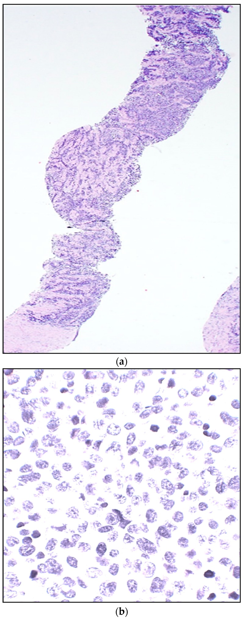
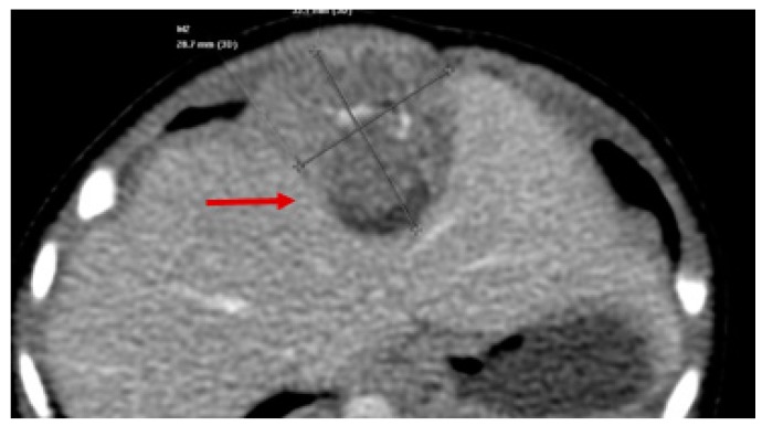
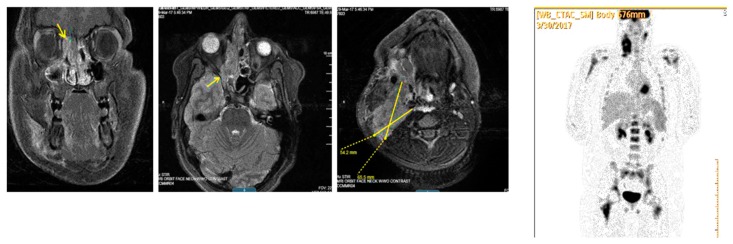
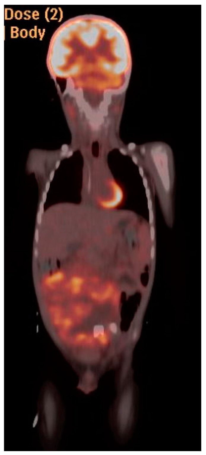
Rhabdomyosarcoma (RMS) is a malignant tumor that represents the most common form of pediatric soft tissue sarcoma. It arises from mesenchymal origin and forms part of the group of small round blue cell tumors of childhood. It has a constant annual incidence of 4.5 cases per 1,000,000 children. The known histological diagnosis of the two major subtypes (embryonal and alveolar) has been recently enhanced by tumor biological markers and molecular differentiation diagnostic tools that have improved not only the updated classification based on risk stratification, but also the treatment approach based on the clinical group. Ewing sarcoma (ES) is a round cell tumor, highly malignant and poorly differentiated that is currently the second most common malignant bone tumor in children. In rare instances, it develops from an extraskeletal origin, classified as extraosseous Ewing sarcoma (EES). We provide an updated, evidence-based and comprehensive review of the molecular diagnosis, clinical and diagnostic approach and a multidisciplinary medical and surgical management according to the latest standard of care for the treatment of pediatric RMS and EES.

Keywords: extraosseous Ewing sarcoma; pediatric; rhabdomyosarcoma.


Conflict of interest statement

The authors declare no conflict of interest.

Figures

Figure 1
Figure 1
(a) Embryonal rhabdomyosarcoma (ERMS)—Botryoid (grape-like) gross appearance. Mucosal (and submucosal) tumor lifts up and folds the vaginal mucosa, from an infant—a classic clinical and gross presentation. (b) Low power histology eminently corresponds with gross botryoid appearance: Squamous non-keratinizing epithelium of vaginal mucosa covers non-demarcated ribbon-like layers of the tumor, which is relatively primitive and cellular superficially (more densely placed nuclei for a blue layer on H&E stain). (c) Persistent tumor post-treatment, with phenotypically “maturing” rhabdomyoblasts (larger cells with a greater amount of cytoplasm) highlighted on the immunohistochemical stain for desmin.
Figure 1
Figure 1
(a) Embryonal rhabdomyosarcoma (ERMS)—Botryoid (grape-like) gross appearance. Mucosal (and submucosal) tumor lifts up and folds the vaginal mucosa, from an infant—a classic clinical and gross presentation. (b) Low power histology eminently corresponds with gross botryoid appearance: Squamous non-keratinizing epithelium of vaginal mucosa covers non-demarcated ribbon-like layers of the tumor, which is relatively primitive and cellular superficially (more densely placed nuclei for a blue layer on H&E stain). (c) Persistent tumor post-treatment, with phenotypically “maturing” rhabdomyoblasts (larger cells with a greater amount of cytoplasm) highlighted on the immunohistochemical stain for desmin.
Figure 2
Figure 2
(a) Alveolar (ARMS)—Needle core biopsy from a soft tissue arm mass from a female teenager. A highly cellular “small round blue cell neoplasm” is seen infiltrating connective tissue in skeletal muscle. (H&E stain). (b) At high power, tumor cells are tightly packed, with variable molding cell membranes, small to moderate amount of delicate cytoplasm, relatively uniform round nuclei, delicate to “salt-and-pepper” chromatin, mostly inconspicuous nucleoli, and scattered mitoses (H&E stain). (c) Immunohistochemical stain for myogenin strongly highlights nuclei of the overwhelming majority of infiltrating tumor cells, densely packed. In contrast, nuclei of the collagenous connective tissue in the background remain appropriately negative.
Figure 2
Figure 2
(a) Alveolar (ARMS)—Needle core biopsy from a soft tissue arm mass from a female teenager. A highly cellular “small round blue cell neoplasm” is seen infiltrating connective tissue in skeletal muscle. (H&E stain). (b) At high power, tumor cells are tightly packed, with variable molding cell membranes, small to moderate amount of delicate cytoplasm, relatively uniform round nuclei, delicate to “salt-and-pepper” chromatin, mostly inconspicuous nucleoli, and scattered mitoses (H&E stain). (c) Immunohistochemical stain for myogenin strongly highlights nuclei of the overwhelming majority of infiltrating tumor cells, densely packed. In contrast, nuclei of the collagenous connective tissue in the background remain appropriately negative.
Figure 3
Figure 3
Axial CT showing Diaphragmatic Rhabdomyosarcoma (red arrow).
Figure 4
Figure 4
MRI (Coronal View) of patient with hepatobiliary embryonal rhabdomyosarcoma (RMS) (red arrow).
Figure 5
Figure 5
Axillary Sentinel Lymph Node Biopsy for trunk RMS. Blue dye can be appreciated in the sample.
Figure 6
Figure 6
PET/CT and MRI of 19 y/o patient with Stage IV extremity alveolar RMS. Uptake can be appreciated in the left axillary node, left elbow and left femoral head.
Figure 7
Figure 7
MRI T-1 weighted image of 13 y/o patient with Stage IV parameningeal alveolar RMS. (Left) image shows in a coronal plane a heterogeneous mass centered within the right ethmoid air cells (arrow) and superior nasal cavity mildly displacing the orbit. (Right) image—axial cut shows right neck lymphadenopathy shown as well with high uptake in PET scan image at the far right.
Figure 8
Figure 8
MRI (sagittal view) of 15 y/o patient with Stage IV perineal alveolar RMS.
Figure 9
Figure 9
Coronal MRI of extraosseous Ewing sarcoma. Note cystic nature of lesion on T2 weighted image.
Figure 10
Figure 10
PET CT coronal view of intraabdominal Ewing sarcoma.

References

    1. Dasgupta R., Fuchs J., Rodeberg D. Rhabdomyosarcoma. Semin. Pediatr. Surg. 2016;25:276–283. doi: 10.1053/j.sempedsurg.2016.09.011. - DOI - PubMed
    1. Ognjanovic S., Linabery A.M., Charbonneau B., Ross J.A. Trends in childhood rhabdomyosarcoma incidence and survival in the United States, 1975–2005. Cancer. 2009;115:4218–4226. doi: 10.1002/cncr.24465. - DOI - PMC - PubMed
    1. Miller R.W., Young J.L., Novakovic B. Childhood cancer. Cancer. 1995;75(Suppl. 1):395–405. doi: 10.1002/1097-0142(19950101)75:1+<395::AID-CNCR2820751321>3.0.CO;2-W. - DOI - PubMed
    1. Rodeberg D., Paidas C. Childhood rhabdomyosarcoma. Semin. Pediatr. Surg. 2006;15:57–62. doi: 10.1053/j.sempedsurg.2005.11.009. - DOI - PubMed
    1. Perez E.A., Kassira N., Cheung M.C., Koniaris L.G., Neville H.L., Sola J.E. Rhabdomyosarcoma in children: A SEER population based study. J. Surg. Res. 2011;170:e243–e251. doi: 10.1016/j.jss.2011.03.001. - DOI - PubMed