Review
. 2018 Dec 10;9(12):617.
doi: 10.3390/genes9120617.

Mechanisms of DNA Methyltransferase Recruitment in Mammals

Affiliations
Review

Mechanisms of DNA Methyltransferase Recruitment in Mammals

Marthe Laisné et al. Genes (Basel). .

Abstract

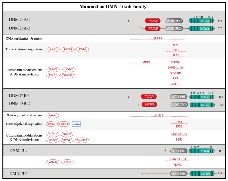
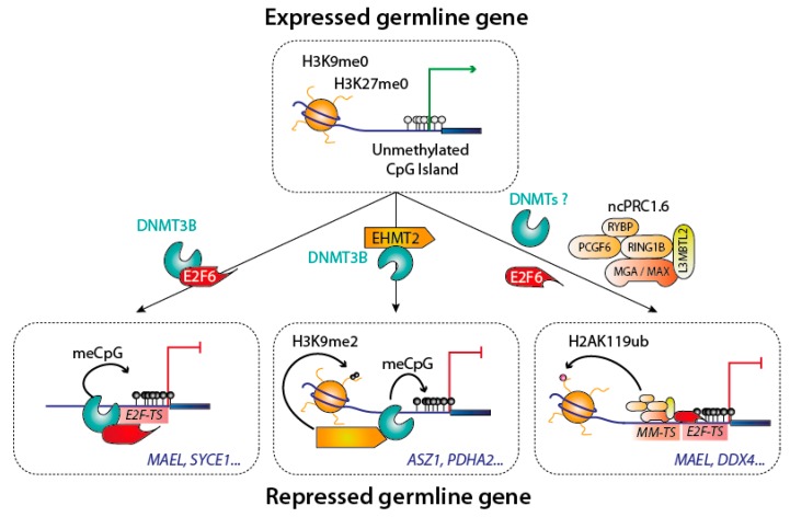
DNA methylation is an essential epigenetic mark in mammals. The proper distribution of this mark depends on accurate deposition and maintenance mechanisms, and underpins its functional role. This, in turn, depends on the precise recruitment and activation of de novo and maintenance DNA methyltransferases (DNMTs). In this review, we discuss mechanisms of recruitment of DNMTs by transcription factors and chromatin modifiers-and by RNA-and place these mechanisms in the context of biologically meaningful epigenetic events. We present hypotheses and speculations for future research, and underline the fundamental and practical benefits of better understanding the mechanisms that govern the recruitment of DNMTs.

Keywords: DNA methylation; DNA methyltransferases; epigenetics.


Conflict of interest statement

The authors declare no conflict of interest.

Figures

Figure 1
Figure 1
Genome-wide distribution of DNA methylation in mammalian species. DNA methylation occurs symmetrically on CpG sites: to simplify, only one DNA strand is shown on this figure. The mammalian genome has a low frequency of CpGs, but the majority of these are methylated (black lollipops) in intergenic regions, repeated elements and transposable elements, and in gene bodies. Conversely, CpG islands are rich in CpGs and are usually protected from DNA methylation (white lollipops). Unmethylated CpG islands frequently correspond to active promoters (green arrow).
Figure 2
Figure 2
Biological processes involving DNA methylation, DNA methyltransferases (DNMTs) and keys partners. DNA methylation is involved in regulating many cellular processes; for all of them, DNMTs cooperate with several essential actors. (a,b) These DNA methylation events can affect the whole genome, as during the two methylation waves during mammalian life cycle, but also after DNA replicates at each cell cycle. (c,d) DNA methylation events can also be localized, as on the newly synthesized DNA after DNA repair. Selected examples, depicted in this cartoon, will be discussed further in this article. Abbreviations used: PGC: Progenitor germ cells; HR: Homologous Recombination; NER: Nucleotide Excision Repair; BER: Base Excision Repair.
Figure 3
Figure 3
Specificities of the DNMTs DNA methylation can take place in two different contexts, and is processed by two distinct mechanisms: de novo DNA methylation involves fully unmethylated DNA and occurs on both strands, whereas maintenance of DNA methylation involves hemimethylated DNA.
Figure 4
Figure 4
Schematic structure of the prokaryotic methyltransferase M. HhaI, compare to DNMT1 and partners. The human DNMT1 contains 1616 amino acids residues. The catalytic methyltransferase domain (MTase, in blue) is very similar to that of the prokaryotic methyltransferase M. HhaI and harbors highly conserved motifs (I-X, in dark blue). In addition, DNMT1 harbors a charge-rich (C-R) domain containing the proliferating cell nuclear antigen (PCNA) binding domain (PBD), an intrinsically disordered domain (IDD) with a nuclear localization sequence (NLS), a replication foci target sequence (RFTS), a zinc finger domain (CXXC), and two bromo-adjacent homology domains (BAH 1/2). The catalytic and the regulatory domains are connected by a series of Gly-Lys repeats. Auto-inhibitory domains are highlighted in green. In addition, some interacting proteins and RNAs are represented: if they are known, mapped interaction domains are indicated. Partners with unknown binding sites are shown on the left. Proteins are depicted in red, RNAs in blue.
Figure 5
Figure 5
Schematic structure of DNMT3 sub-family and partners. Human DNMT3A, DNMT3B, DNMT3L and the rodent DNMT3C contain 912, 953, 387 and 740 amino acids residues, respectively and for the longest isoforms. For DNMT3A and DNMT3B, transcriptional isoforms due to alternative promoters are shown. The catalytic methyltransferase domain (MTase, in blue) harbors highly conserved motifs (I-X, in dark blue). DNMT3A and DNMT3B comprise a PWWP domain and an ATRX-DNMTB-DNMT3L and Plant-Homeodomain (ADD-PHD), that is also found in DNMT3L and DNMT3C. Interacting proteins and RNA are depicted as in Figure 4.
Figure 6
Figure 6
Silencing of the germline genes during implantation. Different complexes are involved in the silencing of germline genes. Three well documented mechanisms are shown, with some example of known targeted germline genes in blue. Abbreviations used: E2F-TS: E2F target sequence; MM-TS: MGA-MAX target sequence.

References

    1. Yadav T., Quivy J.-P., Almouzni G. Chromatin plasticity: A versatile landscape that underlies cell fate and identity. Science. 2018;361:1332–1336. doi: 10.1126/science.aat8950. - DOI - PubMed
    1. Atlasi Y., Stunnenberg H.G. The interplay of epigenetic marks during stem cell differentiation and development. Nat. Rev. Genet. 2017;18:643–658. doi: 10.1038/nrg.2017.57. - DOI - PubMed
    1. Nashun B., Hill P.W.S., Hajkova P. Reprogramming of cell fate: Epigenetic memory and the erasure of memories past. EMBO J. 2015;34:1296–1308. doi: 10.15252/embj.201490649. - DOI - PMC - PubMed
    1. Dawson M.A. The cancer epigenome: Concepts, challenges, and therapeutic opportunities. Science. 2017;355:1147–1152. doi: 10.1126/science.aam7304. - DOI - PubMed
    1. Kungulovski G., Jeltsch A. Epigenome editing: State of the art, concepts, and perspectives. Trends Genet. TIG. 2016;32:101–113. doi: 10.1016/j.tig.2015.12.001. - DOI - PubMed

LinkOut - more resources