. 2018 Dec 8;10(12):1950.
doi: 10.3390/nu10121950.

Oleuropein, the Main Polyphenol of Olea europaea Leaf Extract, Has an Anti-Cancer Effect on Human BRAF Melanoma Cells and Potentiates the Cytotoxicity of Current Chemotherapies

Affiliations

Oleuropein, the Main Polyphenol of Olea europaea Leaf Extract, Has an Anti-Cancer Effect on Human BRAF Melanoma Cells and Potentiates the Cytotoxicity of Current Chemotherapies

Jessica Ruzzolini et al. Nutrients. .

Abstract

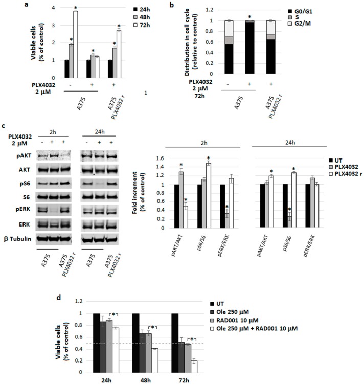
Oleuropein (Ole), a secoiridoid glucoside present in Olea europaea leaves, gained scientific interest thanks to its several biological properties, including the anticancer one. We verified whether Ole might potentiate the cytotoxicity of conventional drugs used to treat melanoma, disclosing a potentially new therapeutic strategy. We tested the cytotoxic action of Ole alone or in combination with chemotherapeutics on A375 human melanoma cells. We found that Ole was able, at a dose of 500 µM, to stimulate apoptosis, while at a non-toxic dose of 250 µM, it affected cell proliferation and induced the downregulation of the pAKT/pS6 pathway. A dose of 250 µM Ole did not potentiate the effect of Vemurafenib (PLX4032), but it succeeded in increasing the cytotoxic effect of Dacarbazine (DTIC). The major effect was found in the association between Ole and Everolimus (RAD001), also on PLX4032-resistant BRAF melanoma cells, which possibly cooperate in the inhibition of the pAKT/pS6 pathway. Of interest, an olive leaf extract enriched in equimolar Ole was more effective and able to further improve DTIC and RAD001 efficacy on BRAF melanoma cells with respect to Ole alone. Therefore, Ole represents a natural product able to potentiate a wide array of chemotherapeutics against BRAF melanoma cells affecting the pAKT/pS6 pathway.

Keywords: BRAF melanoma; Oleuropein; chemotherapeutics; extra virgin oil; olive leaf extract.


Conflict of interest statement

The authors declare no conflict of interest.

Figures

Figure 1
Figure 1
Ole-Vemurafenib (PLX4032) efficacy on A375 melanoma cells. (a) Dose–time response evaluated via MTT assay. (b) (Left) Colony forming units (CFU) assay of alive cells selected using a trypan blue exclusion test after 250 µM (≈125 µg/mL) Ole and/or d treatment for 72 h. (Right) Quantification data of the reduction of colony numbers compared to UT. UT = untreated.
Figure 2
Figure 2
Ole-Dacarbazine (DTIC) efficacy on A375 melanoma cells. (a) Dose–time response evaluated via MTT assay. (b) (Left) Colony Forming Units (CFU) assay of alive cells selected using a trypan blue exclusion test after the treatment with 250 µM (≈125 µg/mL) Ole and/or 270 and 540 µM DTIC (≈50 and 100 µg/mL) for 72 h. (Right) Quantification data of colony numbers compared to UT. (c) (Left) Representative Western blot of PARP1, cleaved PARP1, cleaved caspase 3, pAKT, AKT, pS6, S6, pERK, and ERK after a 250 µM Ole treatment and/or 270 and 540 µM DTIC for 48 h. (Right) Densitometric quantification of the cleaved PARP1, cleaved caspase 3, and of the ratio of pERK/ERK, pAKT/AKT, and pS6/S6 relative to β-tubulin expression, expressed as a fold increment (%) compared to UT. * p ≤ 0.05 refers to Ole-DTIC treatment vs. DTIC alone. UT = untreated.
Figure 3
Figure 3
Ole-Everolimus (RAD001) efficacy on A375 melanoma cells. (a) Dose–time response evaluated via MTT assay. (b) (Left) Colonies forming units (CFU) assay of alive cells selected using a trypan blue exclusion test after a 250 µM (≈125 µg/mL) Ole treatment with and/or 10 and 20 µM RAD001 (≈9.5 and 19 µg/mL) for 48 h. (Right) Quantification data of the colony numbers compared to UT. (c) (Left) Representative Western blot of PARP1, cleaved PARP1, cleaved caspase 3, pAKT, AKT, pS6, S6, pERK, and ERK after 250 µM Ole treatment with and/or 10 and 20 µM RAD001 for 24 h. (Right) Densitometric quantification of the cleaved PARP1, cleaved caspase 3, and of the ratio of pERK/ERK, pAKT/AKT, and pS6/S6 relative to β-tubulin expression, expressed as a fold increment (%) compared to UT. * p ≤ 0.05 refers to Ole-RAD001 treatment vs. RAD001 alone. UT = untreated.
Figure 4
Figure 4
Characterization of A375 melanoma cells resistant to PLX4032 and effect of Ole-RAD001 treatment on these cells. (a) Cell viability evaluated via MTT assay. Significance refers to the untreated control. (b) Cell cycle distribution analyzed using FACS. * p ≤ 0.05 vs. UT. (c) Representative Western blot of pAKT, AKT, pS6, S6, pERK, and ERK in cells treated with PLX4032 2 µM (≈980 ng/mL) for 2 or 24 h. (Right) Densitometric quantification of the ratio of pERK/ERK, pAKT/AKT, and pS6/S6 relative to β-Tubulin expression. * p ≤ 0.05 refers to UT. (d) Dose–time response evaluated on A375 cells that were PLX4032-resistant via MTT assay. * p ≤ 0.05 refers to Ole-RAD001 treatment vs. RAD001 alone. UT = untreated.
Figure 5
Figure 5
Effects of Ole-enriched leaf extract alone or in combination with DTIC, RAD001, or PLX4032 on A375 melanoma cells. (a) Dose–time response evaluated via MTT assay. * p ≤ 0.05 refers to OleRAD001/DTIC treatment vs. RAD001/DTIC alone. (b) (Left) Colonies forming units (CFU) assay of alive cells selected using a trypan blue exclusion test after Ole-enriched leaf extract treatment with and/or RAD001 for 24 h. (Right) Quantification data of colony numbers with respect to UT. * p ≤ 0.05 refers to Ole-DTIC or Ole-RAD001 treatment compared to DTIC or RAD001 alone. UT = untreated.

References

    1. Tsao H., Chin L., Garraway L.A., Fisher D.E. Melanoma: From mutations to medicine. Genes Dev. 2012;26:1131–1155. doi: 10.1101/gad.191999.112. - DOI - PMC - PubMed
    1. Davies H., Bignell G.R., Cox C., Stephens P., Edkins S., Clegg S., Teague J., Woffendin H., Garnett M.J., Bottomley W., et al. Mutations of the BRAF gene in human cancer. Nature. 2002;417:949–954. doi: 10.1038/nature00766. - DOI - PubMed
    1. Garnett M.J., Marais R. Guilty as charged: B-RAF is a human oncogene. Cancer Cell. 2004;6:313–319. doi: 10.1016/j.ccr.2004.09.022. - DOI - PubMed
    1. Houben R., Becker J.C., Kappel A., Terheyden P., Bröcker E.-B., Goetz R., Rapp U.R. Constitutive activation of the Ras-Raf signaling pathway in metastatic melanoma is associated with poor prognosis. J. Carcinog. 2004;3:6. doi: 10.1186/1477-3163-3-6. - DOI - PMC - PubMed
    1. Wan P.T.C., Garnett M.J., Roe S.M., Lee S., Niculescu-Duvaz D., Good V.M., Jones C.M., Marshall C.J., Springer C.J., Barford D., et al. Mechanism of activation of the RAF-ERK signaling pathway by oncogenic mutations of B-RAF. Cell. 2004;116:855–867. doi: 10.1016/S0092-8674(04)00215-6. - DOI - PubMed