. 2018 Dec 9;6(4):52.
doi: 10.3390/proteomes6040052.

Actinobaculum massiliense Proteome Profiled in Polymicrobial Urethral Catheter Biofilms

Affiliations

Actinobaculum massiliense Proteome Profiled in Polymicrobial Urethral Catheter Biofilms

Yanbao Yu et al. Proteomes. .

Abstract

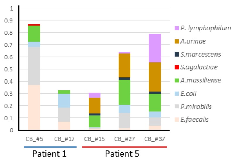
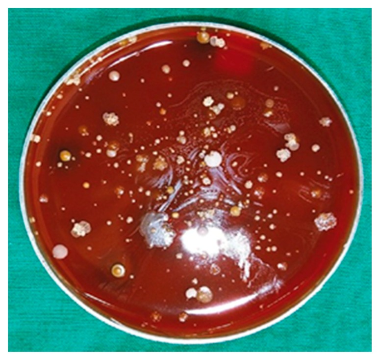
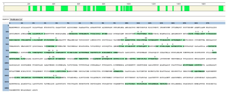
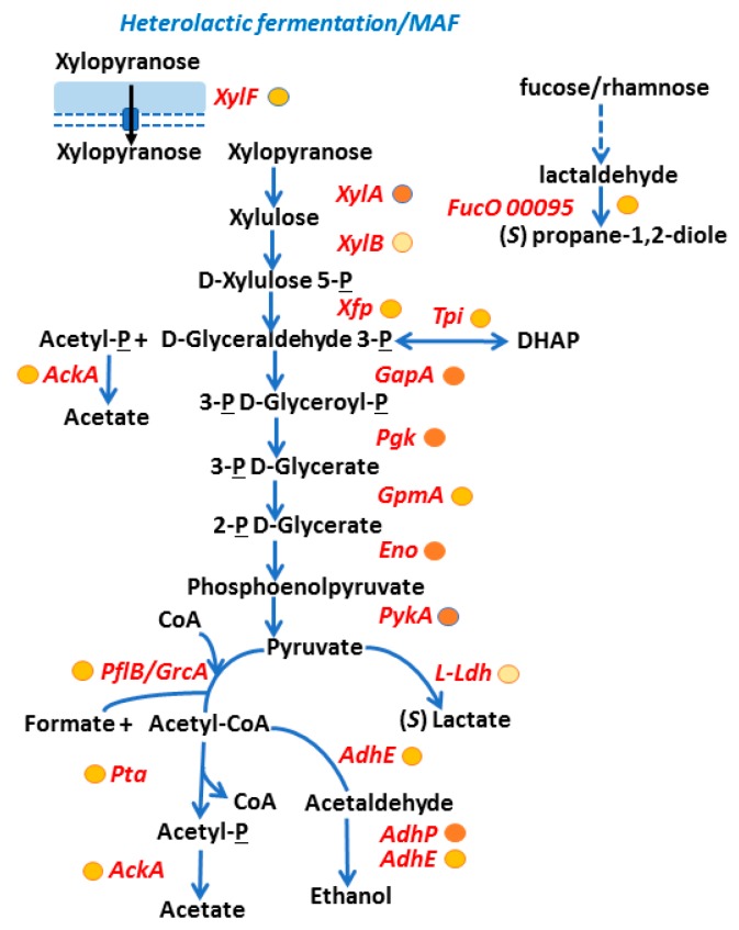
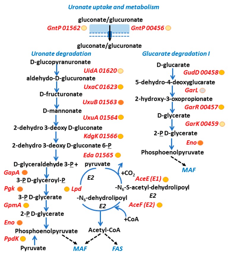
Actinobaculum massiliense, a Gram-positive anaerobic coccoid rod colonizing the human urinary tract, belongs to the taxonomic class of Actinobacteria. We identified A. massiliense as a cohabitant of urethral catheter biofilms (CB). The CBs also harbored more common uropathogens, such as Proteus mirabilis and Aerococcus urinae, supporting the notion that A. massiliense is adapted to a life style in polymicrobial biofilms. We isolated a clinical strain from a blood agar colony and used 16S rRNA gene sequencing and shotgun proteomics to confirm its identity as A. massiliense. We characterized this species by quantitatively comparing the bacterial proteome derived from in vitro growth with that of four clinical samples. The functional relevance of proteins with emphasis on nutrient import and the response to hostile host conditions, showing evidence of neutrophil infiltration, was analyzed. Two putative subtilisin-like proteases and a heme/oligopeptide transporter were abundant in vivo and are likely important for survival and fitness in the biofilm. Proteins facilitating uptake of xylose/glucuronate and oligopeptides, also highly expressed in vivo, may feed metabolites into mixed acid fermentation and peptidolysis pathways, respectively, to generate energy. A polyketide synthase predicted to generate a secondary metabolite that interacts with either the human host or co-colonizing microbes was also identified. The product of the PKS enzyme may contribute to A. massiliense fitness and persistence in the CBs.

Keywords: CAUTI; Keywords: actinobaculum; biofilm; catheter; host-pathogen interaction; metabolism; polymicrobial; proteome; urinary tract infection; uropathogen.


Conflict of interest statement

The authors declare no conflict of interest. The funders had no role in the design of the study; in the collection, analyses, or interpretation of data; in the writing of the manuscript, and in the decision to publish the results.

Figures

Figure 1
Figure 1
Quantitative representation of microbial proteomes in CB samples. The bars are ordered from left to right according to the sequential collection time points. For patient 1, the time points were a week apart; for patient 5, the time points were a month apart. Number gaps do not indicate missed samples. Colored segments of bars represent the relative contribution of a microbial species to the entire proteome. The panel of bacterial species on the right provides the color code for the species as shown in the segmented bar diagram.
Figure 2
Figure 2
Anaerobically grown microorganisms derived from a urethral catheter sample of patient 5 on a blood agar plate. Within 48 hours of growth, various colonies emerged. Among those identified by 16S rRNA analysis on the genus or species level were: Actinobaculum massiliense, Actinomyces sp., Aerococcus sp., Enterococcus sp., Escherichia coli, Finegoldia sp., Morganella morganii, Porphyromonas asaccharolytica, and Prevotella timonensis.
Figure 3
Figure 3
HMPREF9233_01095 protein sequence (a putative subtilisin-like protease). The protein segments from the N- to C-terminus and the peptides identified by shotgun proteomic analysis are highlighted in green (in the bar at the top of the graphic and in amino acid sequence format below, respectively). Modifications are listed above the amino acid position, including deamidation (D) and methionine oxidation (O). These modifications may have occurred during sample processing steps and not reflect biological changes.
Figure 4
Figure 4
Putative A. massiliense peptide uptake, peptide/amino acid metabolism and PKS synthesis functions. The schematic contains protein names in red (short names as annotated in strain ACS-171-V-Col2 database or for orthologs) and/or gene loci (gene locus prefix HMPREF9233_ is not added to the five-digit accession number). The metabolite names are given in black, blue arrows show an enzymatic activity, black arrows a transport activity, and hatched black arrows a cofactor contribution to an enzyme. We provide approximate abundance values of the in vivo detected proteins using circles (behind their names). The darker the fill color, the higher the average abundance level of a protein in averaged in vivo datasets. Proteins predicted to contain cofactors (based on evidence from characterized orthologs) have green symbols underneath/behind the protein names: Me2+ (metal ion), Zn2+ (zinc), py (pyridoxal-5’-phosphate). Other acronyms: ABC, ABC transporter; Lys, Met, Thr, Aro (aromatic acid) BS, enzymes involved in the biosynthesis of amino acids.
Figure 5
Figure 5
Evidence of A. massiliense mixed acid fermentation pathways utilizing xylose in vivo. The protein entities have acronyms/names as explained in the legend of Figure 4. The metabolite names are given in black, blue arrows show an enzymatic activity, black arrows a transport activity, and hatched blue arrows a multi-step metabolic pathway unresolved for this bacterial species. We provide approximate abundance values of in vivo detected proteins using circles (shown behind their names). The darker the fill color, the higher the average abundance level of a protein in in vivo datasets. Acronyms: MAF, mixed acid fermentation; DHAP, dihydroxyacetone phosphate; P, phosphate.
Figure 6
Figure 6
Evidence of active A. massiliense glucuronate and glucarate metabolism pathways in vivo. The protein entities have acronyms/names as explained in the legend of Figure 4. Metabolite names are given in black, blue arrows show an enzymatic activity, black arrows a transport activity, and hatched black arrows a link of a metabolite to a different catabolic pathway. We provide approximate abundance values of in vivo detected proteins using circles (behind their names). The darker the fill color, the higher the average abundance level in vivo. Grey-filled circle: protein not detected in the analyzed A. massiliense proteomes. Acronyms: MAF, mixed acid fermentation; FAS: fatty acid synthesis; P, phosphate; CoA, coenzyme A; E1/E2 cycle, pyruvate dehydrogenase complex.

References

    1. Greub G., Raoult D. "Actinobaculum massiliae," a new species causing chronic urinary tract infection. J. Clin. Microbiol. 2002;40:3938–3941. doi: 10.1128/JCM.40.11.3938-3941.2002. - DOI - PMC - PubMed
    1. Gomez E., Gustafson D.R., Rosenblatt J.E., Patel R. Actinobaculum bacteremia: A report of 12 cases. J. Clin. Microbiol. 2011;49:4311–4313. doi: 10.1128/JCM.00798-11. - DOI - PMC - PubMed
    1. Lawson P.A., Falsen E., Akervall E., Vandamme P., Collins M.D. Characterization of some actinomyces-like isolates from human clinical specimens: Reclassification of Actinomyces suis (soltys and spratling) as Actinobaculum suis comb. Nov. And description of Actinobaculum schaalii sp. Nov. Int. J. Syst. Bacteriol. 1997;47:899–903. doi: 10.1099/00207713-47-3-899. - DOI - PubMed
    1. Cattoir V. Actinobaculum schaalii: Review of an emerging uropathogen. J. Infect. 2012;64:260–267. doi: 10.1016/j.jinf.2011.12.009. - DOI - PubMed
    1. Reinhard M., Prag J., Kemp M., Andresen K., Klemmensen B., Hojlyng N., Sorensen S.H., Christensen J.J. Ten cases of Actinobaculum schaalii infection: Clinical relevance, bacterial identification, and antibiotic susceptibility. J. Clin. Microbiol. 2005;43:5305–5308. doi: 10.1128/JCM.43.10.5305-5308.2005. - DOI - PMC - PubMed