. 2018 Dec 11;23(12):3277.
doi: 10.3390/molecules23123277.

Structural Features of Heparan Sulfate from Multiple Osteochondromas and Chondrosarcomas

Affiliations

Structural Features of Heparan Sulfate from Multiple Osteochondromas and Chondrosarcomas

Noemi Veraldi et al. Molecules. .

Abstract

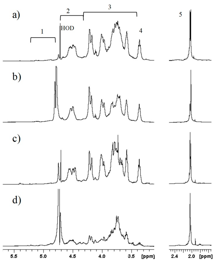
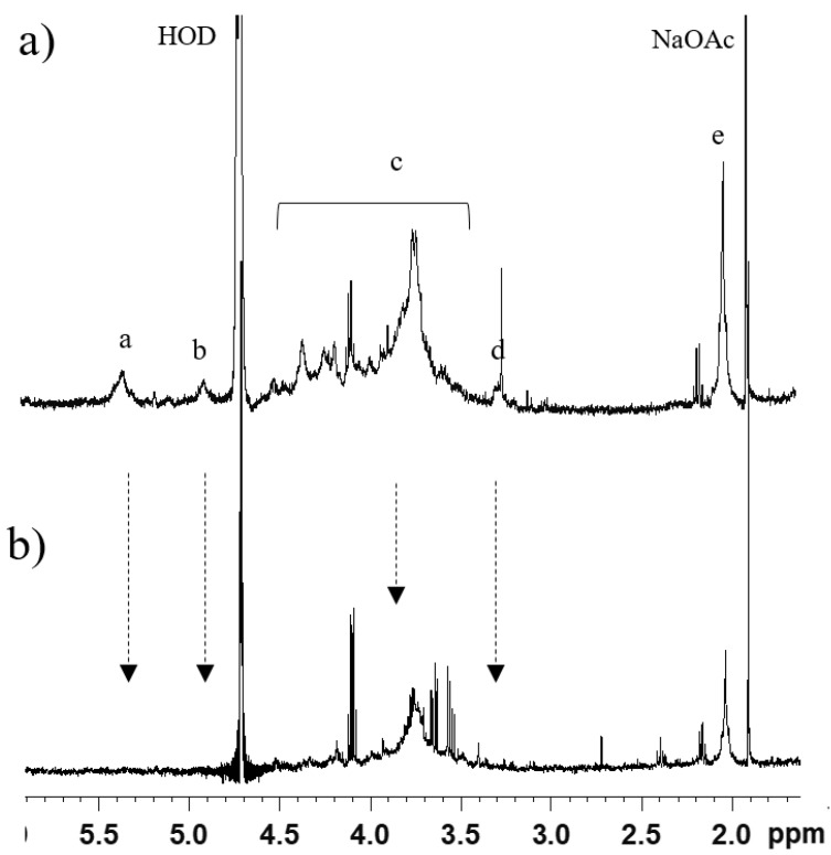
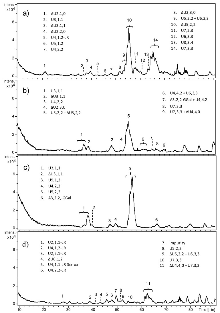
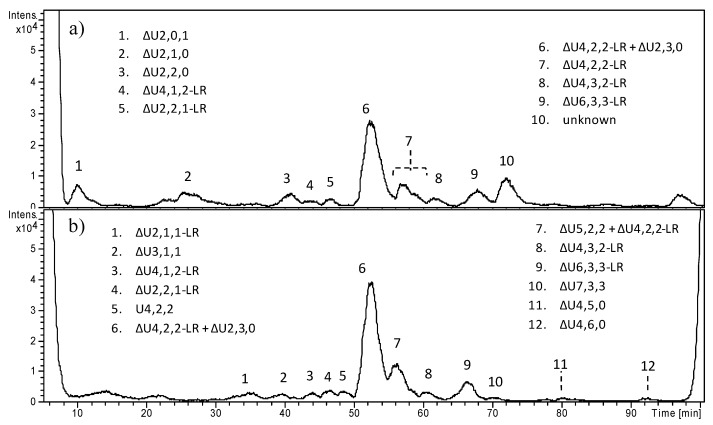
Multiple osteochondromas (MO) is a hereditary disorder associated with benign cartilaginous tumors, known to be characterized by absence or highly reduced amount of heparan sulfate (HS) in the extracellular matrix of growth plate cartilage, which alters proper signaling networks leading to improper bone growth. Although recent studies demonstrated accumulation of HS in the cytoplasm of MO chondrocytes, nothing is known on the structural alterations which prevent HS from undergoing its physiologic pathway. In this work, osteochondroma (OC), peripheral chondrosarcoma, and healthy cartilaginous human samples were processed following a procedure previously set up to structurally characterize and compare HS from pathologic and physiologic conditions, and to examine the phenotypic differences that arise in the presence of either exostosin 1 or 2 (EXT1 or EXT2) mutations. Our data suggest that HS chains from OCs are prevalently below 10 kDa and slightly more sulfated than healthy ones, whereas HS chains from peripheral chondrosarcomas (PCSs) are mostly higher than 10 kDa and remarkably more sulfated than all the other samples. Although deeper investigation is still necessary, the approach here applied pointed out, for the first time, structural differences among OC, PCS, and healthy HS chains extracted from human cartilaginous excisions, and could help in understanding how the structural features of HS are modulated in the presence of pathological situations also involving different tissues.

Keywords: EXT; HPLC–MS; NMR; heparan sulfate; human cartilage; multiple osteochondromas (MO); peripheral chondrosarcoma.


Conflict of interest statement

The authors declare no conflicts of interest.

Figures

Figure 1
Figure 1
Superimposition of 1H-NMR spectra of glycosaminoglycan chains (GAGs) isolated from (a) chondrosarcomas sample 2 (CS2), (b) osteochondromas sample 1 (OC1), (c) prepubescent growth plate samole 1 (GRP1), and (d) fetal growth plate sample 3 (FT3). The main signal regions are indicated: 1, α-anomeric protons; 2, β-anomeric protons; 3, backbone signals; 4, d-glucuronic acid (GlcA) H2; 5, N-acetyl (CH3CO) of N-acetyl-d-glucosamine (GlcNAc). HOD = residual water.
Figure 2
Figure 2
1H-NMR spectra of OC3-B (a) before and (b) after digestion with heparinases. Signals of heparan sulfate (HS) are indicated: a, H-1 of GlcNAc, N-sulfo-d-glucosamine (GlcNS), and 2-O-sulfo-l-iduronic acid (IdoA2S) (5.4 ppm); b, H-5 of IdoA (4.9–5 ppm); c, backbone signals; d, H-2 of GlcA and GlcNS (3.4 ppm); e, N-acetyl (CH3CO) of GlcNAc (2.0 ppm). Arrows indicate the decrease in signal.
Figure 3
Figure 3
LC–MS profiles of heparinase digestion products from fetal HS: (a) FT1, (b) FT2, (c) FT3, and (d) FT4. Oligosaccharides were identified based on their mass/charge ratio (m/z) and labeled as follows: Δ indicates the unsaturated bond of the terminal uronic acid, followed by the number of monomers, sulfate groups, and acetyl groups. When a uronic acid (or a glucosamine) is present at both the reducing end (RE) and non-reducing end (NRE), it is indicated by U (or A). LR indicates the tetrasaccharide G-Gal2-Xyl of the linkage region. LR-Ser-ox indicates the presence of the linkage region bearing an oxidized serine residue.
Figure 4
Figure 4
LC–MS profiles of heparinase digestion products from prepubescent healthy HS: (a) GRP2-A > 10 kDa, and (b) GRP2-B < 10 kDa. Oligosaccharides were identified by their mass/charge ratio (m/z) and labeled as follows: when a uronic acid (or a glucosamine) is present at both the RE and NRE, it is indicated by U (or A). LR indicates the tetrasaccharide linkage region G-Gal2-Xyl. Oligosaccharides for which an imprecise interpretation was found are labeled as “unknown”.
Figure 5
Figure 5
Mass spectra of selected oligosaccharides observed by ion-pair reversed-phase (IPRP) HPLC/electrospray ionization quadrupole time of flight (ESI-Q-TOF). On the left, the (a) experimental and (b,c) theoretical isotope patterns of a selected oligosaccharide together with the possible interpretations are reported. On the right, the (d) experimental and (e) theoretical isotope patterns of the standard trisulfated disaccharide are reported for comparison.
Figure 6
Figure 6
LC–MS profiles of heparinase digestion of HS from osteochondromas: (a) OC4-A, (b) OC4-B, (c) OC5-A, and (d) OC5-B. When a uronic acid (or a glucosamine) is present at both the RE and NRE, it is indicated by U (or A). LR indicates the tetrasaccharide G-Gal2-Xyl of the linkage region, while LR-Ser-ox indicates also the presence of the oxidized serine residue. Oligosaccharides for which an imprecise interpretation was found were labeled as “unknown”.
Figure 7
Figure 7
LC–MS profiles of heparinase digestion of HS from chondrosarcoma: (a) CS1-A, (b) CS1-B, (c) CS3-A, and (d) CS3-B. For some m/z ratios, more than one oligosaccharide structure was possible. When a uronic acid (or a glucosamine) is present at both the RE and NRE, it is indicated by U (or A). LR indicates the tetrasaccharide linkage region G-Gal2-Xyl. Oligosaccharides for which an imprecise interpretation was found were labeled as “unknown”.

References

    1. Bernfield M., Gotte M., Park P.W., Reizes O., Fitzgerald M.L., Lincecum J., Zako M. Functions of cell surface heparan sulfate proteoglycans. Annu. Rev. Biochem. 1999;68:729–777. doi: 10.1146/annurev.biochem.68.1.729. - DOI - PubMed
    1. Bishop J.R., Schuksz M., Esko J.D. Heparan sulphate proteoglycans fine-tune mammalian physiology. Nature. 2007;446:1030–1037. doi: 10.1038/nature05817. - DOI - PubMed
    1. Sarrazin S., Lamanna W.C., Esko J.D. Heparan sulfate proteoglycans. Cold Spring Harb. Perspect Biol. 2011;3 doi: 10.1101/cshperspect.a004952. - DOI - PMC - PubMed
    1. Lin X. Functions of heparan sulfate proteoglycans in cell signaling during development. Development. 2004;131:6009–6021. doi: 10.1242/dev.01522. - DOI - PubMed
    1. Esko J.D., Selleck S.B. Order out of chaos: Assembly of ligand binding sites in heparan sulfate. Annu. Rev. Biochem. 2002;71:435–471. doi: 10.1146/annurev.biochem.71.110601.135458. - DOI - PubMed

MeSH terms