. 2018 Dec 12;23(12):3302.
doi: 10.3390/molecules23123302.

Fucoidan Exerts Anticancer Effects Against Head and Neck Squamous Cell Carcinoma In Vitro

Affiliations

Fucoidan Exerts Anticancer Effects Against Head and Neck Squamous Cell Carcinoma In Vitro

Wiktoria Blaszczak et al. Molecules. .

Abstract

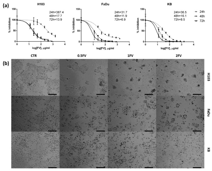
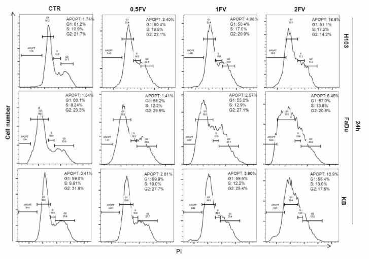
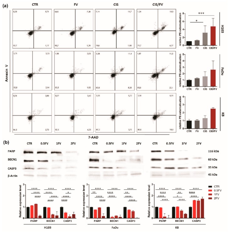
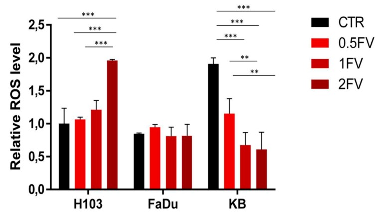
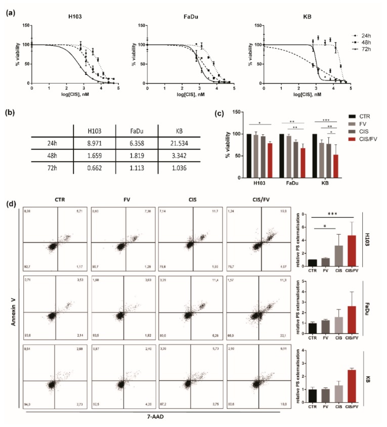
Fucoidans have been reported to exert anticancer effects with simultaneous low toxicity against healthy tissue. That correlation was observed in several cancer models, however, it has never been investigated in head and neck cancer before. To magnify the efficacy of conventional therapy, the administration of agents like fucoidan could be beneficial. The aim of this study was to evaluate the anticancer effect of Fucus vesiculosus (FV) extract alone and with co-administration of cisplatin in head and neck squamous cell carcinoma (HNSCC) in vitro. MTT assay results revealed an FV-induced inhibition of proliferation in all tested cell lines (H103, FaDu, KB). Flow cytometric cell cycle analysis showed an FV-induced, dose-dependent arrest in either S/G2 phase (H103, FaDu) or G1 arrest (KB). Furthermore, a dose-dependent gain in apoptotic fraction was observed. Western blot analysis confirmed the induction of apoptosis. A significant dose-dependent increase in reactive oxygen species (ROS) production was revealed in the H103 cell line, while FaDu cells remained unresponsive. On the contrary, an HPV-positive cell line, KB, demonstrated a dose-dependent decrease in ROS synthesis. Moreover, fucoidan enhanced the response to cisplatin (synergistic effect) in all cell lines with the HPV-positive one (KB) being the most sensitive. These results have been confirmed by flow-cytometric apoptosis analysis. In conclusion, we confirmed that fucoidan exhibits anticancer properties against HNSCC, which are manifested by the induction of apoptosis, regulation of ROS production, cell cycle arrest, and inhibition of proliferation.

Keywords: HNSCC; complementary therapy; fucoidan; head and neck cancer; marine algae.


Conflict of interest statement

The authors declare no conflict of interest. The funders had no role in the design of the study; in the collection, analyses, or interpretation of data; in the writing of the manuscript, or in the decision to publish the results.

Figures

Figure 1
Figure 1
Effect of fucoidan on the proliferation and viability of HNSCC cell lines: H103, FaDu, KB. (a) Inhibition of proliferation of tested cell lines after incubation with fucoidan for 24, 48 and 72 h. The data are presented as a mean ± standard deviation. (b) The decreased proliferation of tested cell lines after 24 h incubation with different concentrations of fucoidan was observed (CTR—control, 0.5FV—half-fold of IC50 concentration, 1FV—IC50 concentration, 2FV—2-fold of IC50 concentration). Pictures were taken under 40× magnification. Scale bars represent 200 µm.
Figure 2
Figure 2
Impact of fucoidan on cell cycle distribution of HNSCC cell lines: H103, FaDu, KB. Cell cycle analysis after incubation with different concentrations of fucoidan for 24 h. Increasing dose of fucoidan increases the number of apoptotic cells in all tested cell lines. Treatment with fucoidan induces G2/S arrest in H103 and FaDu cells, and G1 arrest in KB cell line. (CTR—control, 0.5FV—half-fold of IC50 concentration, 1FV—IC50 concentration, 2FV—2-fold of IC50 concentration, PI—propidium iodide).
Figure 3
Figure 3
Impact of fucoidan on the induction of apoptosis in HNSCC cell lines: H103, FaDu, KB (a): 24 h incubation with fucoidan induces apoptosis in all tested cell lines (CTR—control, 0.5FV—half-fold of IC50 concentration, 1FV—IC50 concentration, 2FV—2-fold of IC50 concentration, PS—phosphatidylserine). The data are presented as an average ± standard deviation. (b): 24 h treatment with fucoidan down-regulates the levels of full-length PARP, BECN1, and CASP3 in H103 and FaDu cells. Fucoidan decreases the expression of PARP and BECN1 while up-regulating levels of CASP3 in KB cell line. (CTR—control, 0.5FV—half-fold of IC50 concentration, 1FV—IC50 concentration, 2FV—2-fold of IC50 concentration). The data are presented as mean ± standard deviation. p values lower than 0.05 were considered significant and are labeled by asterisks (*) for p < 0.05, (**) for p < 0.01, (***) for p < 0.001, and (****) for p ≤ 0.0001.
Figure 4
Figure 4
Impact of fucoidan on the synthesis of reactive oxygen species in HNSCC cells. Twenty-four hour treatment with fucoidan up-regulates ROS production in a dose-dependent manner in H103 cells, whilst down-regulating it in KB cells. No effect was observed on FaDu cells. (CTR—control, 0.5FV—half-fold of IC50 concentration, 1FV—IC50 concentration, 2FV—2-fold of IC50 concentration). The data are presented as an average ± standard deviation.
Figure 5
Figure 5
Simultaneous co-administration of cisplatin and fucoidan on HNSCC cell lines: H103, FaDu, KB. (a) Survival curves of HNSCC cells generated after exposure to cisplatin for 24, 48 and 72 h. (b): IC50 doses after 24, 48 and 72 h incubation with cisplatin in µM. (c) Fucoidan enhances the response to cisplatin by further decreasing the proliferation of all tested cell lines after 8 h incubation (H103) and 16 h (FaDu, KB) at the IC50 concentrations. The data are presented as mean ± standard deviation. (d) Fucoidan enhances the response to cisplatin by further inducing apoptosis of all tested cell lines after 8 h incubation (H103) and 16 h (FaDu, KB) at the IC50 concentrations. CTR—control, CIS—cisplatin, CIS/FV—cisplatin and fucoidan, PS—phosphatidylserine). p values lower than 0.05 were considered significant and are labelled by asterisks (*) for p < 0.05, (**) for p < 0.01, (***) for p < 0.001, and (****) for p ≤ 0.0001.

References

    1. Berteau O., Mulloy B. Sulfated fucans, fresh perspectives: Structures, functions, and biological properties of sulfated fucans and an overview of enzymes active toward this class of polysaccharide. Glycobiology. 2003;13:29–40. doi: 10.1093/glycob/cwg058. - DOI - PubMed
    1. Blaszczak W., Sobecka A., Barczak W., Suchorska W.M. Fucoidan and its potential as a complementary agent in cancer therapy. Lett. Oncol. Sci. 2017;14:86–95.
    1. Sanjeewa K.K.A., Lee J.S., Kim W.S., Jeon Y.J. The potential of brown-algae polysaccharides for the development of anticancer agents: An update on anticancer effects reported for fucoidan and laminaran. Carbohydr. Polym. 2017;177:451–459. doi: 10.1016/j.carbpol.2017.09.005. - DOI - PubMed
    1. Han M.H., Lee D.S., Jeong J.W., Hong S.H., Choi I.W., Cha H.J., Kim S., Kim H.S., Park C., Kim G.Y., et al. Fucoidan Induces ROS-Dependent Apoptosis in 5637 Human Bladder Cancer Cells by Downregulating Telomerase Activity via Inactivation of the PI3K/Akt Signaling Pathway. Drug Dev. Res. 2016;78:37–48. doi: 10.1002/ddr.21367. - DOI - PubMed
    1. Liu F., Luo G., Xiao Q., Chen L., Luo X., Lv J., Chen L. Fucoidan inhibits angiogenesis induced by multiple myeloma cells. Oncol. Rep. 2016;36:1963–1972. doi: 10.3892/or.2016.4987. - DOI - PubMed

MeSH terms

LinkOut - more resources