Comparative Study
. 2018 Dec 13;19(1):27.
doi: 10.1186/s12860-018-0178-8.

Comparative evaluation of mesenchymal stromal cells from umbilical cord and amniotic membrane in xeno-free conditions

Affiliations
Comparative Study

Comparative evaluation of mesenchymal stromal cells from umbilical cord and amniotic membrane in xeno-free conditions

Yongxu Mu et al. BMC Cell Biol. .

Abstract

Background: Within the past years, umbilical cord (UC) and amniotic membrane (AM) expanded in human platelet lysate (PL) have been found to become increasingly candidate of mesenchymal stromal cells (MSCs) in preclinical and clinical studies. Different sources of MSCs have different properties, and lead to different therapeutic applications. However, the similarity and differences between the AMMSCs and UCMSCs in PL remain unclear.

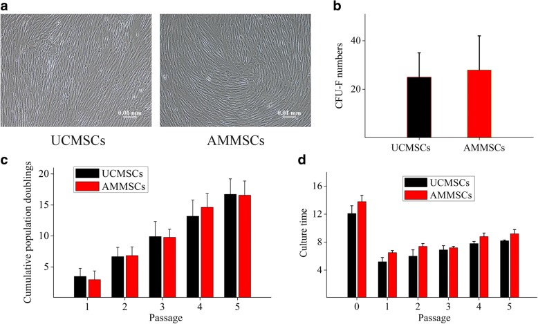
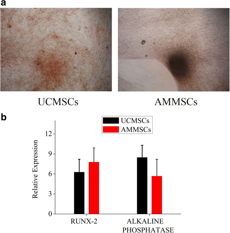
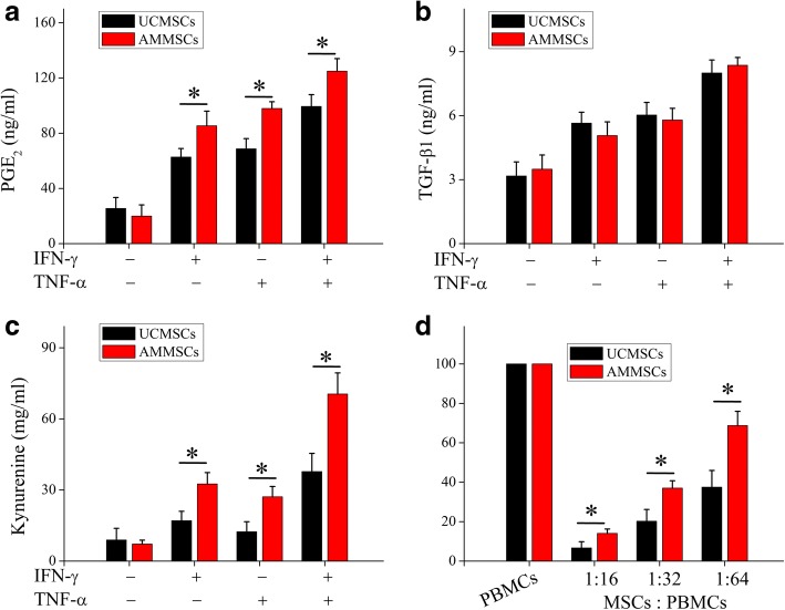
Results: In this study, we conduct a direct head-to-head comparison with regard to biological characteristics (morphology, immunophenotype, self-renewal capacity, and trilineage differentiation potential) and immunosuppression effects of AMMSCs and UCMSCs expanded in PL. Our results indicated that AMMSCs showed similar morphology, immunophenotype, proliferative capacity and colony efficiency with UCMSCs. Moreover, no significantly differences in osteogenic, chondrogenic and adipogenic differentiation potential were observed between the two types of cells. However, AMMSCs exhibited higher PGE2 expression and IDO activity compared with UCMSCs when primed by IFN-γ and (or) TNF-α induction, and AMMSCs showed a higher inhibitory effect on PBMCs proliferation than UCMSCs.

Conclusion: The results suggest that AMMSCs expanded in PL showed similar morphology, immunophenotype, self-renewal capacity, and trilineage differentiation potential with UCMSCs. However, AMMSCs possessed superior immunosuppression effects in comparison with UCMSCs. These results suggest that AMMSCs in PL might be more suitable than UCMSCs for treatment of immune diseases. This work provides a novel insight into choosing the appropriate source of MSCs for treatment of immune diseases.

Keywords: Amnion; Characteristics; Immunomodulatory; Mesenchymal stromal cells; Platelet lysate; Umbilical cord.


Conflict of interest statement

Ethics approval and consent to participate

All human samples were obtained from healthy donors that provided an informed, written consent for research use, and the study was approved by the Ethics Committee of the First Affiliated Hospital of Baotou Medical College.

Consent for publication

Not applicable.

Competing interests

The authors declare that they have no competing interests.

Publisher’s Note

Springer Nature remains neutral with regard to jurisdictional claims in published maps and institutional affiliations.

Figures

Fig. 1
Fig. 1
Platelet lysis released sufficient amounts of growth factors in PL. a Schematic overview of preparation of PL. PRP was generated from peripheral blood through a series of centrifugations. Based on platelet counts (109 platelets/ml) after the centrifugation step, sufficient PPP was removed to achieve PRP. PL was prepared from PRP by a simple freeze-thawing procedure. b The quantification of growth factors in PL and PPP was determined using ELISA. Bars represented means ± SD. n = 5; *p < 0.05, **p < 0.01, and ***p < 0.005
Fig. 2
Fig. 2
AMMSCs in PL showed similar morphology and self-renewal capacity with UCMSCs. a Both UCMSCs and ATMSCs expanded in PL showed fibroblast-like morphologies. Scale bar: 10 μm. b Clone-forming ability of UCMSCs and AMMSCs was assessed by CFU-F counts per 1 × 105 TNCs. The proliferation capacity of UCMSCs and AMMSCs was assessed by CPD c and culture time d of each passage. Bars represented means ± SD; n = 5
Fig. 3
Fig. 3
AMMSCs in PL showed similar osteogenic differentiation potential with UCMSCs. a The osteogenic differentiation of AMMSCs and UCMSCs was confirmed by Alizarin Red staining of mineralization. Magnification × 100. b Comparative investigation of osteogenic differentiation capability of UCMSCs and AMMSCs was assessed by quantitative analysis of RUNX-2 and alkaline phosphatase mRNA expression. Bars represented means ± SD; n = 5
Fig. 4
Fig. 4
AMMSCs in PL showed similar chondrogenic differentiation potential with UCMSCs. a The chondrogenic differentiation of AMMSCs and UCMSCs was confirmed by Alcian Blue staining of glycosaminoglycans. Magnification × 100. b Comparative investigation of chondrogenic differentiation capability of UCMSCs and AMMSCs was assessed by quantitative analysis of SOX-9 and collagen II mRNA expression. Bars represented means ± SD; n = 5
Fig. 5
Fig. 5
AMMSCs in PL showed similar adipogenic differentiation potential with UCMSCs. a The adipogenic differentiation of AMMSCs and UCMSCs was confirmed by Oil Red O staining of lipid vacuoles. Magnification × 100. b Comparative investigation of adipogenic differentiation capability of UCMSCs and AMMSCs was assessed by quantitative analysis of PPARg and LPL mRNA expression. Bars represented means ± SD; n = 5
Fig. 6
Fig. 6
AMMSCs in PL showed superior immunosuppression effects with UCMSCs. When primed by IFN-γ and/or TNF-α induction, PGE2 (a) and TGF-β1 (b) expression were analyzed. IDO activity (c) was evaluated by kynurenine levels. (d) UCMSCs suppressed allogeneic lymphocyte proliferation. Bars represented means ± SD, n = 5; *P < 0.05

References

    1. Squillaro T, Peluso G, Galderisi U. Clinical trials with mesenchymal stem cells: an update. Cell Transplant. 2016;25:829–848. doi: 10.3727/096368915X689622. - DOI - PubMed
    1. Solomon J, Csontos L, Clarke D, Bonyhadi M, Zylberberg C, McNiece I, et al. Current perspectives on the use of ancillary materials for the manufacture of cellular therapies. Cytotherapy. 2016;18:1–12. doi: 10.1016/j.jcyt.2015.09.010. - DOI - PubMed
    1. Capelli C, Domenghini M, Borleri G, Bellavita P, Poma R, Carobbio A, et al. Human platelet lysate allows expansion and clinical grade production of mesenchymal stromal cells from small samples of bone marrow aspirates or marrow filter washouts. Bone Marrow Transplant. 2007;40:785–791. doi: 10.1038/sj.bmt.1705798. - DOI - PubMed
    1. Hemeda H, Giebel B, Wagner W. Evaluation of human platelet lysate versus fetal bovine serum for culture of mesenchymal stromal cells. Cytotherapy. 2014;16:170–180. doi: 10.1016/j.jcyt.2013.11.004. - DOI - PubMed
    1. Sharma RR, Pollock K, Hubel A, McKenna D. Mesenchymal stem or stromal cells: a review of clinical applications and manufacturing practices. Transfusion. 2014;54:1418–1437. doi: 10.1111/trf.12421. - DOI - PMC - PubMed

Publication types

Substances