Comparative Study
. 2018 Dec 18;72(24):3112-3122.
doi: 10.1016/j.jacc.2018.09.073.

His Resynchronization Versus Biventricular Pacing in Patients With Heart Failure and Left Bundle Branch Block

Affiliations
Comparative Study

His Resynchronization Versus Biventricular Pacing in Patients With Heart Failure and Left Bundle Branch Block

Ahran D Arnold et al. J Am Coll Cardiol. .

Abstract

Background: His bundle pacing is a new method for delivering cardiac resynchronization therapy (CRT).

Objectives: The authors performed a head-to-head, high-precision, acute crossover comparison between His bundle pacing and conventional biventricular CRT, measuring effects on ventricular activation and acute hemodynamic function.

Methods: Patients with heart failure and left bundle branch block referred for conventional biventricular CRT were recruited. Using noninvasive epicardial electrocardiographic imaging, the authors identified patients in whom His bundle pacing shortened left ventricular activation time. In these patients, the authors compared the hemodynamic effects of His bundle pacing against biventricular pacing using a high-multiple repeated alternation protocol to minimize the effect of noise, as well as comparing effects on ventricular activation.

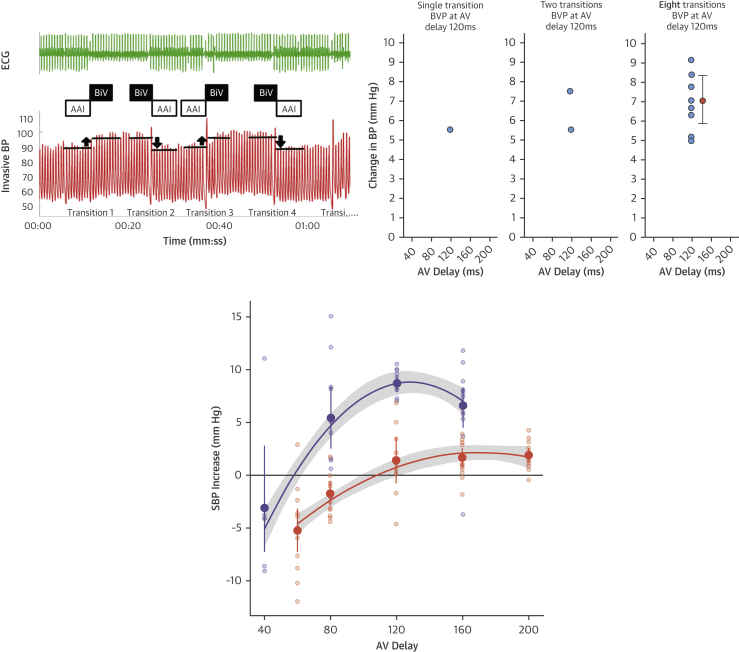
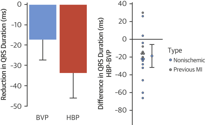
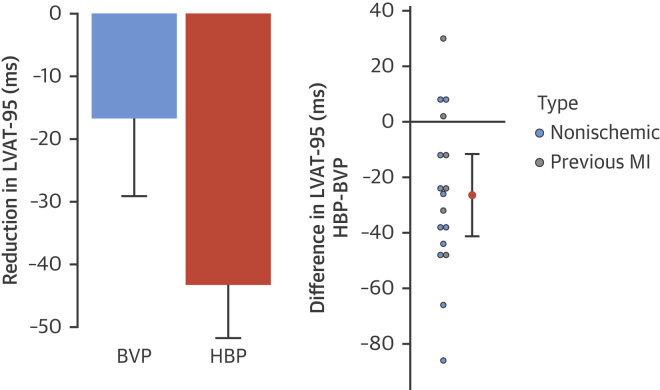
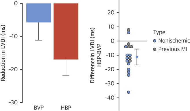
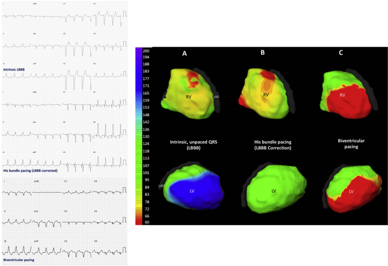
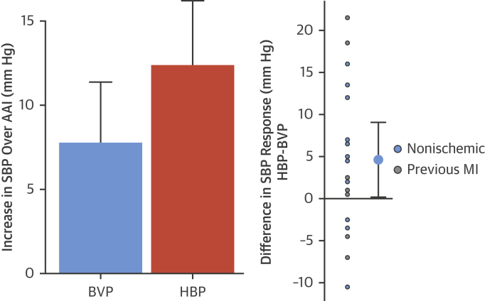
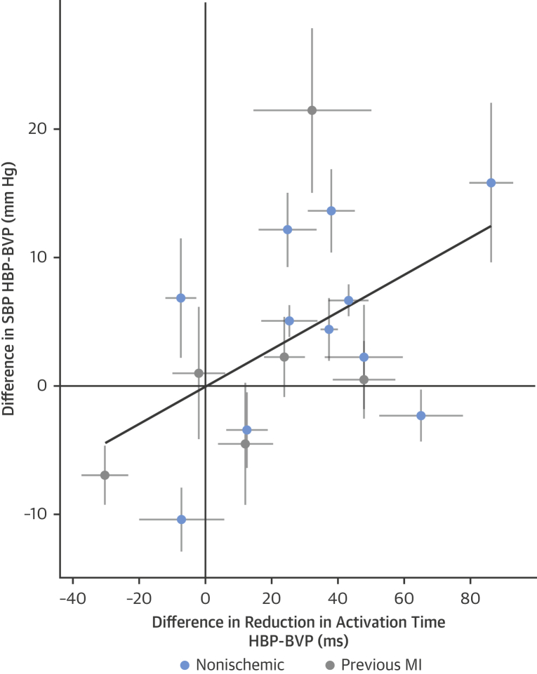
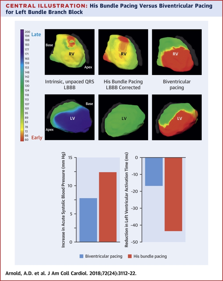
Results: In 18 of 23 patients, left ventricular activation time was significantly shortened by His bundle pacing. Seventeen patients had a complete electromechanical dataset. In them, His bundle pacing was more effective at delivering ventricular resynchronization than biventricular pacing: greater reduction in QRS duration (-18.6 ms; 95% confidence interval [CI]: -31.6 to -5.7 ms; p = 0.007), left ventricular activation time (-26 ms; 95% CI: -41 to -21 ms; p = 0.002), and left ventricular dyssynchrony index (-11.2 ms; 95% CI: -16.8 to -5.6 ms; p < 0.001). His bundle pacing also produced a greater acute hemodynamic response (4.6 mm Hg; 95% CI: 0.2 to 9.1 mm Hg; p = 0.04). The incremental activation time reduction with His bundle pacing over biventricular pacing correlated with the incremental hemodynamic improvement with His bundle pacing over biventricular pacing (R = 0.70; p = 0.04).

Conclusions: His resynchronization delivers better ventricular resynchronization, and greater improvement in hemodynamic parameters, than biventricular pacing.

Keywords: ECGI; His bundle pacing; His resynchronization therapy; biventricular pacing; cardiac resynchronization therapy; noninvasive epicardial mapping.


Figures

None
Graphical abstract
Figure 1
Figure 1
Analysis of Hemodynamic Data (Top left) A minimum of 4 alternations between AAI pacing and HBP or BVP were performed for each tested AV delay (therefore, a total of 8 transitions). For each alternation, relative change in SBP was calculated and the mean of the 8 transitions was calculated. (Top right) As more transitions occur, more data points are collected for change in SBP, eventually resulting in 10 values with mean and confidence intervals. Single or few measurements result in highly inaccurate estimates due to variability. (Bottom) A mean and confidence interval are calculated for each AV delay for both HBP (purple) and BVP (orange). An example is shown of quadratic curves fitted to data from multiple transitions at a range of AV delays to produce the peak systolic blood pressure responses. AV = atrioventricular; BP = blood pressure; BVP = biventricular pacing; ECG = electrocardiogram; HBP = His bundle pacing; SBP = systolic blood pressure.
Figure 2
Figure 2
12-Lead Surface ECG QRS Responses Change in QRS duration with BVP and HBP (left) and within-patient incremental QRS duration reduction with HBP over BVP (right). The 95% confidence intervals are displayed. MI = myocardial infarction; other abbreviations as in Figure 1.
Figure 3
Figure 3
LVAT-95 Responses Change in LVAT-95 with BVP and HBP (left) and within-patient incremental reduction of LVAT-95 with HBP over BVP (right). The 95% confidence intervals are displayed. LVAT-95 = left ventricular activation time spanning 95% of activations; other abbreviations as in Figures 1 and 2.
Figure 4
Figure 4
LVDI Responses Change in LVDI with BVP and HBP (left) and within-patient incremental reduction of LVDI with HBP over BVP (right). The 95% confidence intervals are displayed. LVDI = left ventricular dyssynchrony index; other abbreviations as in Figures 1 and 2.
Figure 5
Figure 5
12-Lead Surface ECGs and ECGI Activation Maps (Left) 12-lead surface ECGs of intrinsic LBBB, His bundle pacing correction of LBBB, and biventricular pacing in a single patient. (Right) Noninvasive ECGI epicardial maps of LV and RV activation recorded from the same patient during (A) intrinsic rhythm (LBBB), (B) selective His bundle pacing, and (C) biventricular pacing. The color scale (left) shows that the late (blue) activation that occurs on the lateral wall of the LV during intrinsic activation is not seen with His bundle pacing: the LBBB pattern of activation is no longer present during His bundle pacing; a normal physiological ventricular pattern is seen instead. The small regions of red, early activation on the RV during his bundle pacing, may represent subtle nonselectivity of capture or interpolation of signal noise misidentified as activation. During biventricular pacing, activation spreads from early activation sites in both the RV and LV. ECG = electrocardiogram; ECGI = electrocardiographic imaging; LAD = left anterior descending artery; LBBB = left bundle branch block; LV = left ventricle/ventricular; RV = right ventricle/ventricular.
Figure 6
Figure 6
Hemodynamic Responses Acute improvement in systolic blood pressure is observed with both BVP and HBP (left). His bundle pacing delivered significantly greater within-patient improvements in acute systolic blood pressure (right). The 95% confidence intervals are displayed. Abbreviations as in Figures 1 and 2.
Figure 7
Figure 7
Correlation Between Electrical and Hemodynamic Responses Within-patient comparison of the difference in left ventricular activation time with HBP compared with BVP and the difference in acute SBP. Thin black lines = 95% confidence intervals in both axes. Thick black line = the regression line. Abbreviations as in Figures 1 and 2.
Central Illustration
His Bundle Pacing Versus Biventricular Pacing for Left Bundle Branch Block (Top) Noninvasive epicardial RV and LV activation maps of intrinsic LBBB, His bundle pacing (HBP), and biventricular pacing (BVP). HBP produces a more physiological activation pattern than BVP. (Bottom) HBP produces a greater increase in acute systolic blood pressure and greater reduction in LV activation time than BVP. LBBB = left bundle branch block; LV = left ventricular; RV = right ventricular.

Comment in

References

    1. Sohaib S., Chen Z., Whinnett Z.I. Meta-analysis of symptomatic response attributable to the pacing component of cardiac resynchronization therapy. Eur J Heart Fail. 2013;15:1419–1428. - PubMed
    1. Cleland J.G., Abraham W.T., Linde C. An individual patient meta-analysis of five randomized trials assessing the effects of cardiac resynchronization therapy on morbidity and mortality in patients with symptomatic heart failure. Eur Heart J. 2013;34:3547–3556. - PMC - PubMed
    1. Leclercq C., Cazeau S., Le Breton H. Acute hemodynamic effects of biventricular DDD pacing in patients with end-stage heart failure. J Am Coll Cardiol. 1998;32:1825–1831. - PubMed
    1. Cleland J.G., Daubert J.-C., Erdmann E. The effect of cardiac resynchronization on morbidity and mortality in heart failure. N Engl J Med. 2005;352:1539–1549. - PubMed
    1. Jones S., Lumens J., Sohaib S.A., BRAVO Investigators Cardiac resynchronization therapy: mechanisms of action and scope for further improvement in cardiac function. Europace. 2017;19:1178–1186. - PMC - PubMed

Publication types