. 2019 Jan 4;363(6422):84-87.
doi: 10.1126/science.aav6740. Epub 2018 Dec 13.

Structure of the posttranslational Sec protein-translocation channel complex from yeast

Affiliations

Structure of the posttranslational Sec protein-translocation channel complex from yeast

Samuel Itskanov et al. Science. .

Abstract

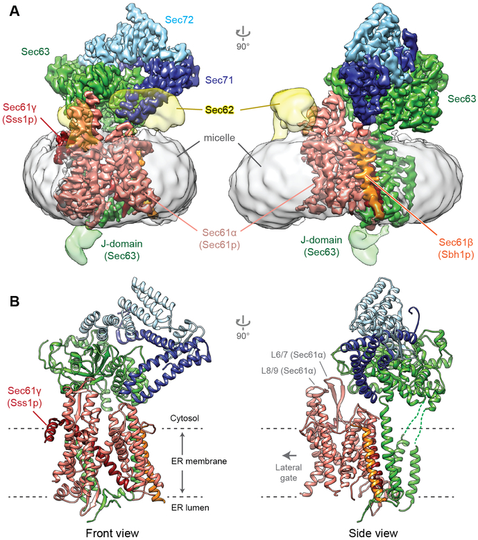
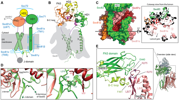
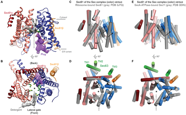
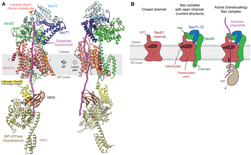
The Sec61 protein-conducting channel mediates transport of many proteins, such as secretory proteins, across the endoplasmic reticulum (ER) membrane during or after translation. Posttranslational transport is enabled by two additional membrane proteins associated with the channel, Sec63 and Sec62, but its mechanism is poorly understood. We determined a structure of the Sec complex (Sec61-Sec63-Sec71-Sec72) from Saccharomyces cerevisiae by cryo-electron microscopy (cryo-EM). The structure shows that Sec63 tightly associates with Sec61 through interactions in cytosolic, transmembrane, and ER-luminal domains, prying open Sec61's lateral gate and translocation pore and thus activating the channel for substrate engagement. Furthermore, Sec63 optimally positions binding sites for cytosolic and luminal chaperones in the complex to enable efficient polypeptide translocation. Our study provides mechanistic insights into eukaryotic posttranslational protein translocation.


Figures

Figure 1.
Figure 1.. Structure of the yeast Sec complex.
Cryo-EM density map (A) and atomic model (B) of the yeast posttranslational protein translocation complex. Front view, view into the lateral gate.
Figure 2.
Figure 2.. Structure of Sec63 and interactions with the channel.
(A) A schematic of Sec63 domains. Regions interacting with other parts of the complex are indicated by blue lines. Unmodeled regions are shown in dashed lines. (B) Structure of Sec63 (front view). The position of Sec61 is shown by a gray shade. (C) Interactions between TMs of Sec63 and Sec61. Left, a view from the back; right, a cutaway view from the ER lumen. Black arrowed line, the cross-sectional plane. Note that TMs 2, 9, and 10 of Sec61α are located above the cross-sectional plane. (D) Interactions between Sec63 and Sec61 in the luminal side. Left, a β-sheet formed between Sec61α (TM5 indicated by a dashed line) and the segment between Sec63 TM3 and the J-domain. Right, a magnified view with side chains in sticks. (E) Interactions between the FN3 domain and the cytosolic loop L6/7 of Sec61α (also see Fig. 1B).
Figure 3.
Figure 3.. A fully opened Sec61 channel in the Sec complex.
(A and B) Structure of the Sec61 channel. The N- and C- terminal halves of Sec61α are in blue and salmon, respectively. Gray density feature is presumed detergent molecules. Pore-lining residues are shown as green balls and sticks. Density feature for the plug is in purple. ‘2’ and ‘7’ indicate TM2 and TM7 respectively. (CF) Comparison of Sec61 of the Sec complex (colored) with Sec61 of the cotranslational ribosome-Sec61 complex (gray; C and D) or SecY of a bacterial posttranslational SecA-SecY channel complex (gray; E and F). The structures are aligned with respect to the C-terminal half of Sec61α (C–F). Shown are the front (A, C, and E) and cytosolic (B, D, and F) views. Numbers indicate corresponding TMs. Dashed line, lateral gate. Asterisk, translocation pore. For simplicity, L6/7 and L8/9 of Sec61α were not shown. In D and F, TMs of Sec63 are also shown (green). Also see fig. S6 for comparisons to archaeal SecY and substrate-engaged channels.
Figure 4.
Figure 4.. Model of an active translocation complex.
(A) The Sec complex structure superimposed with a Ssa1p C-terminal peptide (red orange; PDB ID: 5L0Y) and DnaK Hsp70 as a model for BiP (yellow and brown; PDB ID: 5RNO). (B) Schematics for a closed Sec61 channel in isolation (left), an open channel in association with Sec63 (middle), and an active Sec complex engaged with a substrate (right; corresponding to the model in (A)). For the full translocation cycle, see fig S8.

Comment in

References

    1. Park E, Rapoport TA, Mechanisms of Sec61/SecY-mediated protein translocation across membranes. Annu Rev Biophys 41, 21–40 (2012). - PubMed
    1. Voorhees RM, Hegde RS, Toward a structural understanding of co-translational protein translocation. Curr Opin Cell Biol 41, 91–99 (2016). - PubMed
    1. Mandon EC, Trueman SF, Gilmore R, Protein translocation across the rough endoplasmic reticulum. Cold Spring Harb Perspect Biol 5, (2013). - PMC - PubMed
    1. Van den Berg B et al., X-ray structure of a protein-conducting channel. Nature 427, 36–44 (2004). - PubMed
    1. Rothblatt JA, Deshaies RJ, Sanders SL, Daum G, Schekman R, Multiple genes are required for proper insertion of secretory proteins into the endoplasmic reticulum in yeast. J Cell Biol 109, 2641–2652 (1989). - PMC - PubMed

Publication types

MeSH terms

Substances