. 2018 Dec 26;115(52):E12255-E12264.
doi: 10.1073/pnas.1814810115. Epub 2018 Dec 13.

TANGO1 and SEC12 are copackaged with procollagen I to facilitate the generation of large COPII carriers

Affiliations

TANGO1 and SEC12 are copackaged with procollagen I to facilitate the generation of large COPII carriers

Lin Yuan et al. Proc Natl Acad Sci U S A. .

Abstract

Large coat protein complex II (COPII)-coated vesicles serve to convey the large cargo procollagen I (PC1) from the endoplasmic reticulum (ER). The link between large cargo in the lumen of the ER and modulation of the COPII machinery remains unresolved. TANGO1 is required for PC secretion and interacts with PC and COPII on opposite sides of the ER membrane, but evidence suggests that TANGO1 is retained in the ER, and not included in normal size (<100 nm) COPII vesicles. Here we show that TANGO1 is exported out of the ER in large COPII-coated PC1 carriers, and retrieved back to the ER by the retrograde coat, COPI, mediated by the C-terminal RDEL retrieval sequence of HSP47. TANGO1 is known to target the COPII initiation factor SEC12 to ER exit sites through an interacting protein, cTAGE5. SEC12 is important for the growth of COPII vesicles, but it is not sorted into small budded vesicles. We found both cTAGE5 and SEC12 were exported with TANGO1 in large COPII carriers. In contrast to its exclusion from small transport vesicles, SEC12 was particularly enriched around ER membranes and large COPII carriers that contained PC1. We constructed a split GFP system to recapitulate the targeting of SEC12 to PC1 via the luminal domain of TANGO1. The minimal targeting system enriched SEC12 around PC1 and generated large PC1 carriers. We conclude that TANGO1, cTAGE5, and SEC12 are copacked with PC1 into COPII carriers to increase the size of COPII, thus ensuring the capture of large cargo.

Keywords: COPII; SEC12; TANGO1; collagen; secretion.


Conflict of interest statement

The authors declare no conflict of interest.

Figures

Fig. 1.
Fig. 1.
TANGO1 is copackaged with PC1 into large COPII carriers. (A) Schematic representation of TANGO1’s domain structure with information on relevant interactions. The N-terminal SH3 domain interacts with the PC-specific chaperone HSP47, which binds to folded PCs (16). The coiled-coil (CC) domains of TANGO1 form a stable complex with cTAGE5 and SEC12 (–19). The PRD of TANGO1 interacts with the COPII inner coat protein SEC23 (14, 20). (B) Scheme depicting the isolation of COPII carriers from cell-free budding reactions as previously described (11, 22). Briefly, COPII vesicles were generated by incubating a reaction containing donor membrane prepared from IMR-90 cells, purified recombinant COPII proteins (1 μg of SAR1B wild-type or H79G, 1 μg of SEC23A/24D, 1 μg of SEC13/31A), nucleotides, and 2 μg/μL HT1080 cytosol at 30 °C for 1 h. Vesicles in 7,000 × g supernatant fractions from budding reactions were isolated by flotation. (C) TANGO1, cTAGE5, and SEC12 in COPII carriers were detected by immunoblotting aliquots of the top float fractions of reactions conducted under different conditions. Donor membrane (DM) was included as an input control. PC1 and HSP47 are captured into large COPII-coated PC1 carriers and serve as positive controls for large COPII carriers. ERGIC53 and SEC22B are found in conventional COPII vesicles and serve as further controls for COPII vesicles. Ribophorin I is an ER resident protein that serves as a negative control. (D) Dual color 3D-STORM images of a KI6 cell containing multiple large COPII coated vesicles. Overview of the cell is shown in (i), and Insets are enlarged and shown in (iixiii). TANGO1 (magenta) colocalized with KLHL12-FLAG (green) which coats large COPII vesicles in xy maximum projections (ii, iii, vi, vii, x, xi) or respective xz cross-sections (iv, v, viii, ix, xii, xiii). (Scale bars: 5 μm in D, i; 500 nm in D, iixiii.)
Fig. 2.
Fig. 2.
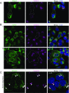
TANGO1 is retrieved with HSP47 via its c-terminal RDEL motif. Confocal microscopy images of U-2OS cells transfected with StrepII-HSP47ΔRDEL. (A and B) Immunofluorescent labeling using antibodies against the StrepII tag (A; green) or HSP47 (B; green) and TANGO1 (magenta). In wild-type cells, TANGO1 localized at ERES and HSP47 localized in the ER (Fig. 3A). TANGO1 mis-localization is observed following the overexpression and secretion of StrepII-HSP47ΔRDEL in cells marked inside dotted lines. In these cells, HSP47 (green) no longer shows ER localization, but rather appeared to localize at the cell surface; little TANGO1 (magenta) is detected in most cells (A and B, i), and localizes to the Golgi membrane in some cells (B, ii). (C) Cells transfected with StrepII-HSP47ΔRDEL were incubated at 19.5 °C in the presence of ascorbate for 3 h to accumulate cargo in the Golgi and followed by immunofluorescence labeling targeting of TANGO1 (green) and a Golgi marker, Golgin97 (magenta). Arrowheads point to examples of TANGO1 colocalized with Golgin97. (Scale bars, 10 μm.)
Fig. 3.
Fig. 3.
TANGO1 localizes around giant COPII membranes in cells depleted of COPI. (A) Confocal images of U-2os-wt-c11 cells transfected with negative control siRNA or siRNA that targeted coatomer subunit δ (ARCN1 gene) for 48 h followed by immunofluorescence labeling of TANGO1 (magenta) and HSP47 (green). TANGO1 was observed around large HSP47 puncta in cells depleted of COPI. Magnified Insets show two examples of such structures. (BD) Confocal images of U-2os-wt-c11 cells expressing ARF1 Q71L GFP (cyan) were labeled by immunofluorescence targeting of TANGO1 (yellow) and HSP47 (magenta) in B or PC1 (magenta) in C or SEC31A (magenta) in D. (Scale bars: 10 μm in overviews and 1 μm in magnified Insets.)
Fig. 4.
Fig. 4.
SEC12 is enriched in large COPII-coated PC1 carriers. (A) Schematic representation of the fractionation procedure used to separate small and large COPII carriers. Supernatant after 7,000 × g centrifugation from a vesicle-budding reaction was overlaid onto a step gradient consisting of 7.5% and 18% OptiPrep. The gradient was centrifuged at 250,000 × g for 1 h to separate large from regular cargo-containing COPII carriers. Fractions (numbered 1–5) taken after sedimentation were used as inputs for flotation to separate vesicles from soluble contents. (B) Analysis of the large cargo PC1 and regular COPII cargos ERGIC53 and SEC22B in buoyant membrane collected from sedimentation fractions postflotation. (C) Budding reactions were supplemented with wild-type or H79G mutant SAR1B. Buoyant membrane from relevant sedimentation fractions were immunoblotted for TANGO1, cTAGE5, and SEC12. PC1 and HSP47 serve as markers for COPII-coated PC1 carriers. ERGIC53 and SEC22B serve as markers for regular COPII vesicles. (D) Confocal image of KI6 cells that were induced for KLHL12 overexpression for 7.5 h and immunolabeled with a fluorescent antibody against SEC31A (green) and SEC12 (magenta). Inset of the merged image is magnified 5× (Lower Right). (Scale bars, 10 μm.) (E) Confocal immunofluorescence images of SEC12 (cyan), PC1 (yellow), and SEC16A (magenta) in Saos-2 cells. Arrowheads point to examples of large SEC12 puncta that colocalized with PC1 but not SEC16A. (Scale bars, 10 μm.)
Fig. 5.
Fig. 5.
SEC12 is localized around PC1 throughout large COPII vesicle formation. (A) Three-dimensional (3D) STORM images of single color large SEC12 (cyan) puncta collected from Saos-2 cells. Three representative examples from three classes of ultrastructures (iiii) are shown in magnified maximum xy projection (Left), virtual cross-sections in xz (Center), and yz (Right). (B) Three-color 3D STORM images of large COPII structures in KI6 cells following 7.5-h induced overexpression of KLHL12: SEC12 (cyan), PC1 (magenta), and COPII coat subunit SEC31A (green in B, i and iii) or KLHL12 (green in B, ii). Representative examples of three classes of ultrastructures (iiii) are shown in three-color merged maximum xy projection (Left), three-color merged virtual cross-sections (Center), and two-color merged virtual cross-sections of SEC12 (cyan) and PC1 (magenta) (Right). (C) Schematic illustrations of three classes of ultrastructures of SEC12 arranged in putative time progression: (i) enrichment of SEC12 (cyan) around PC1 (magenta) containing ER; (ii) nascent large COPII (green) budding event where SEC12 localizes around membrane-containing PC1; (iii) free large COPII-coated PC1 carrier with enriched SEC12. (Scale bars, 500 nm.)
Fig. 6.
Fig. 6.
Active targeting of SEC12 to large cargo increases COPII size. (A) Drawings that represent the working model of SEC12 enrichment (Left) and the design of split GFP constructs (Right). In our working model, the luminal SH3 domain of TANGO1 targets SEC12 to PC1. To recapitulate sorting of SEC12 to PC1, we constructed 3xFLAG-SEC12-GFP11 and TANGO1-lumi-HA-GFP1-10 fusion constructs, where TANGO1-lumi contains TANGO1’s cargo-sensing SH3 domain. (B) GFP complementation brought SEC12 and TANGO1-lumi together. Confocal images of U-2OS-wt-c11 cells that were transfected with TANGO1-lumi-HA-GFP1-10 alone (Top), 3xFLAG-SEC12-GFP11 alone (Middle), or both (Bottom), complemented GFP (cyan), TANGO1-lumi-HA-GFP1-10 (yellow, IF against HA), and 3xFLAG-SEC12-GFP11 (magenta, IF against FLAG). (CE) Confocal images of U-2OS-wt-c11 cells transfected with both TANGO1-lumi-HA-GFP1-10 and 3xFLAG-SEC12-GFP11 Cells were immunofluorescently labeled against PC1 (magenta) in C and E and imaged in combination with complemented GFP (green). (D and E) Cells were cultured in dialyzed medium, which contained minimal ascorbate. Ascorbate was added to cells for 30 min to stimulate PC1 export. Confocal z-stacks of magnified Insets show hollow spherical complement GFP (green) structures that entirely enveloped PC1 (magenta). Confocal z-stack was taken with a step size of 0.38 μm. [Scale bars: 10 μm (BE) and 1 μm (magnified Insets in D and E).]

References

    1. Dancourt J, Barlowe C. Protein sorting receptors in the early secretory pathway. Annu Rev Biochem. 2010;79:777–802. - PubMed
    1. Barlowe C, et al. COPII: A membrane coat formed by Sec proteins that drive vesicle budding from the endoplasmic reticulum. Cell. 1994;77:895–907. - PubMed
    1. Futai E, Hamamoto S, Orci L, Schekman R. GTP/GDP exchange by Sec12p enables COPII vesicle bud formation on synthetic liposomes. EMBO J. 2004;23:4146–4155. - PMC - PubMed
    1. Lee MC, et al. Sar1p N-terminal helix initiates membrane curvature and completes the fission of a COPII vesicle. Cell. 2005;122:605–617. - PubMed
    1. Antonny B, Madden D, Hamamoto S, Orci L, Schekman R. Dynamics of the COPII coat with GTP and stable analogues. Nat Cell Biol. 2001;3:531–537. - PubMed

Publication types

MeSH terms