Review
. 2018 Dec 17;46(6):1753-1770.
doi: 10.1042/BST20180004. Epub 2018 Dec 13.

Structure, activation and dysregulation of fibroblast growth factor receptor kinases: perspectives for clinical targeting

Affiliations
Review

Structure, activation and dysregulation of fibroblast growth factor receptor kinases: perspectives for clinical targeting

Brendan Farrell et al. Biochem Soc Trans. .

Abstract

The receptor tyrosine kinase family of fibroblast growth factor receptors (FGFRs) play crucial roles in embryonic development, metabolism, tissue homeostasis and wound repair via stimulation of intracellular signalling cascades. As a consequence of FGFRs' influence on cell growth, proliferation and differentiation, FGFR signalling is frequently dysregulated in a host of human cancers, variously by means of overexpression, somatic point mutations and gene fusion events. Dysregulation of FGFRs is also the underlying cause of many developmental dysplasias such as hypochondroplasia and achondroplasia. Accordingly, FGFRs are attractive pharmaceutical targets, and multiple clinical trials are in progress for the treatment of various FGFR aberrations. To effectively target dysregulated receptors, a structural and mechanistic understanding of FGFR activation and regulation is required. Here, we review some of the key research findings from the last couple of decades and summarise the strategies being explored for therapeutic intervention.

Keywords: drug discovery and design; fibroblast growth factor receptors; receptor tyrosine kinases; structural biology.


Conflict of interest statement

The Authors declare that there are no competing interests associated with the manuscript.

Figures

Figure 1.
Figure 1.. Stimulation of FGFRs.
FGFRs are composed of an extracellular domain comprising D1, acid box, and D2 and D3 domains, followed by a single helix TMD, the JMD, and an intracellular ‘split’ tyrosine KD. Two models describing receptor stimulation by FGF ligand and heparin/heparan sulfate cofactor have been described: the canonical ligand-induced receptor dimerisation model (left) and an allosteric ligand-induced conformational change model (right). Receptor activation leads to trans-autophosphorylation of the kinase domains and stimulation of intracellular signalling cascades. The boxed regions (A–C) correspond to those in Figure 2.
Figure 2.
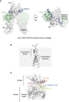
Figure 2.. Structures of FGFR extracellular, transmembrane, and kinase domains.
(A) Crystal structure of FGFR1 extracellular domains D2 and D3 (grey cartoon on transparent surface representation) in a 2:2:2 complex with FGF2 (light green, cartoon) and heparin (dark blue, sticks) (PDB: 1FQ9). Only one copy of FGF2 and FGFR1 are shown in cartoon representation for clarity. (B) An FGFR3 transmembrane domain dimer derived from NMR (PDB: 2LZL) in cartoon representation with the observed dimerisation interface and GxxxG-like motifs highlighted. (C) FGFR3 kinase domain crystal structure (PDB: 4K33) in cartoon representation on a transparent surface with the N- and C-lobes and structural elements, the αC helix (salmon), the P-loop (orange), the catalytic loop (blue), the A-loop (yellow), the kinase hinge (magenta) and the (incomplete) kinase insert (black) highlighted. Panels are not in scale with one another.
Figure 3.
Figure 3.. Comparison of active and inactive FGFR kinase domain states.
(A) Structural overlay (right) of non-phosphorylated, inactive FGFR2 kinase domain (light grey) (PDB: 2PSQ) and phosphorylated, active FGFR2 kinase domain (blue) (PDB: 2PVF), both in cartoon representation. Additionally, in the active kinase domain, the kinase regulatory spine and two participating residues, H624 of the HRD motif and F645 of the DFG motif, are highlighted in red sticks and surface representation. During kinase activation, the molecular brake hydrogen bonding network between H544, N549, E565, and K641 of FGFR2 is broken, as illustrated in the expanded sections (left). The same regions in the inactive state of FGFR1 kinase (PDB: 4V01) (dark grey) with the corresponding H541, N546, E562, and K638 residues are also presented (far left), illustrating the conservation of this feature among FGFRs. (B) Structural differences in A-loop conformation in active and inactive FGFR2 kinase domains where phosphorylation-dependent salt bridge interactions between R649 and phospho-Y657 (pY657) stabilise an extended conformation of the loop in the activated kinase.
Figure 4.
Figure 4.. Point mutations of FGFR3.
The locations of a selection of developmental disease and cancer-associated point mutations of FGFR3 in the extracellular domain (left) (PDB: 1FQ9), the transmembrane domain (middle) (PDB: 2LZL), and the kinase domain (right) (PDB: 4K33), as discussed in the text. As no FGFR3 ligand-dimerised extracellular domain structure is available, the extracellular domain of FGFR1 in complex with FGF2 is shown, illustrating the localisation of FGFR3 point mutations to regions which could generate similar dimer structures in a ligand-independent manner. Similarly, in the kinase domain, the αC helix (salmon), the αEF helix (cyan), the hinge region (magenta), and the A-loop (yellow) are highlighted to illustrate the localisation of many point mutations to important regulatory elements of the kinase domain.
Figure 5.
Figure 5.. Binding modes of FGFR inhibitors.
Crystal structures of inhibitor-bound FGFR1 and FGFR4 kinase domains, illustrating the binding modes of reversible and irreversible (covalent) inhibitors. Reversible inhibitors can be classified into type I and type II inhibitors, differing in their binding modes (top). Type I inhibitors such as AZD4547 bind to active, DFG in-state kinases, whereas type II inhibitors such as ponatinib bind to inactive, DFG out-state kinases. In each instance, FGFR1 kinase domains are shown in full in cartoon representation with transparent surfaces (light grey) and inhibitors in stick representation (purple). Additionally, the inhibitor-binding site is expanded for each with FGFR1 (light grey) in cartoon representation alone, and F642 of the DFG motif (red) and the gatekeeper residue V550 (orange) shown in stick representation with transparent surfaces. In the ponatinib-bound structure, the asterisk (*) indicates the location of the ethynyl group attributed to the ability of ponatinib to accommodate gatekeeper residue mutations and to the multikinase selectivity profile of the inhibitor. The binding modes of three irreversible, covalent inhibitors to FGFR4 and FGFR4 surrogate kinase domain (FGFR1–Y563C) are presented in expanded panels in a similar manner (bottom). In these, where resolved in the crystal structures, the gatekeeper residue and Phe of the DFG motif is shown as above, and the Cys residues utilised in ligand conjugation are highlighted also (yellow). In the FIIN-2-bound structure, F631 (DFG motif, FGFR4) is observed in both the DFG in- and DFG out-states, marked with a double asterisk (**). The structures presented are: FGFR1 kinase domain bound to AZD4547 (PDB: 4V05) and ponatinib (PDB: 4V01); FGFR4 kinase domain bound to BLU-9931 (PDB: 4XCU) and FIIN-2 (PDB: 4QQC) and of FGFR4 surrogate kinase domain (FGFR1 harbouring a Y563C substitution) bound to H3B-6527 (PDB: 5VND).

References

    1. Manning G., Whyte D.B., Martinez R., Hunter T. and Sudarsanam S. (2002) The protein kinase complement of the human genome. Science 298, 1912–1934 10.1126/science.1075762 - DOI - PubMed
    1. Lemmon M.A. and Schlessinger J. (2010) Cell signaling by receptor tyrosine kinases. Cell 141, 1117–1134 10.1016/j.cell.2010.06.011 - DOI - PMC - PubMed
    1. Turner N. and Grose R. (2010) Fibroblast growth factor signalling: from development to cancer. Nat. Rev. Cancer. 10, 116–129 10.1038/nrc2780 - DOI - PubMed
    1. Beenken A. and Mohammadi M. (2009) The FGF family: biology, pathophysiology and therapy. Nat. Rev. Drug Discov. 8, 235–253 10.1038/nrd2792 - DOI - PMC - PubMed
    1. Hart A.W., Baeza N., Apelqvist Å. and Edlund H. (2000) Attenuation of FGF signalling in mouse β-cells leads to diabetes. Nature 408, 864–868 10.1038/35048589 - DOI - PubMed

Publication types

MeSH terms

Substances