. 2019 Feb 1;294(5):1618-1631.
doi: 10.1074/jbc.RA118.005222. Epub 2018 Dec 13.

The trans-Golgi network is a major site for α-secretase processing of amyloid precursor protein in primary neurons

Affiliations

The trans-Golgi network is a major site for α-secretase processing of amyloid precursor protein in primary neurons

Jing Zhi A Tan et al. J Biol Chem. .

Abstract

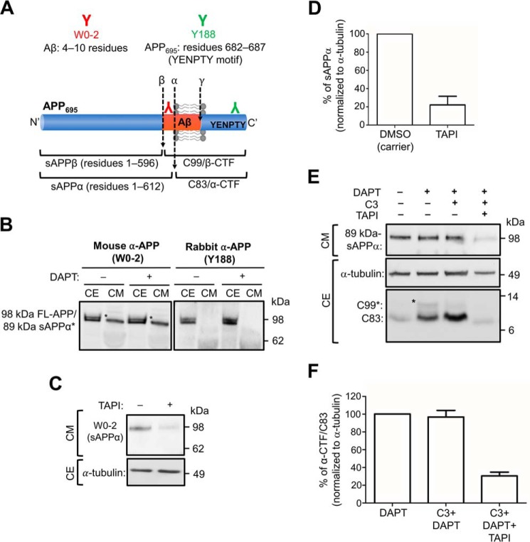
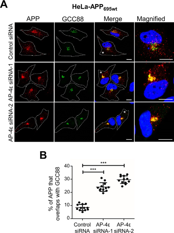
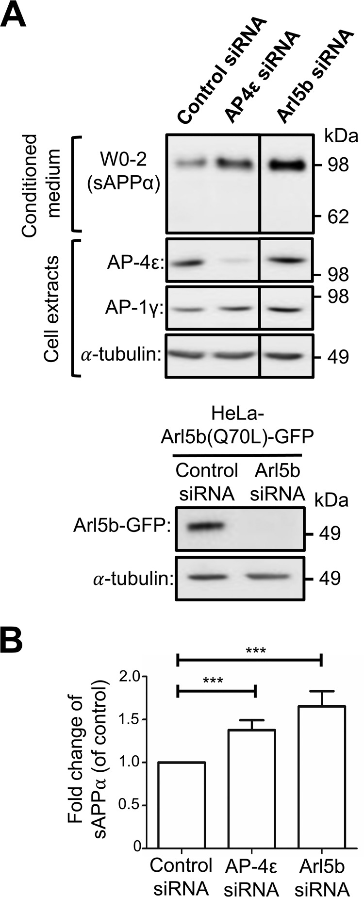
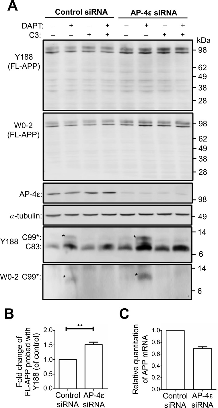
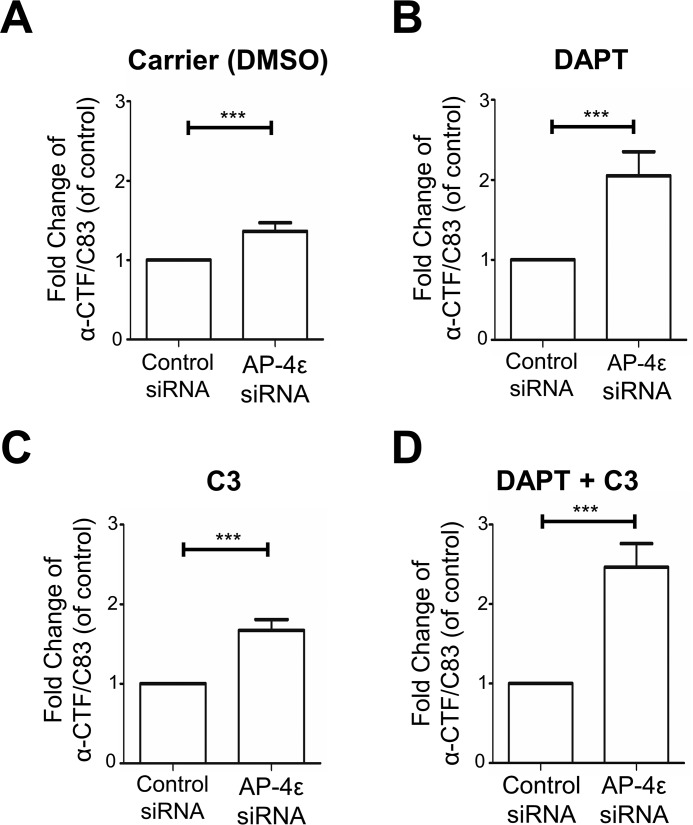
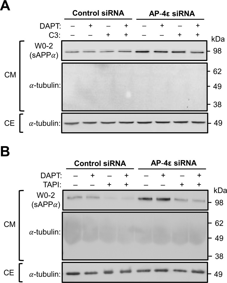
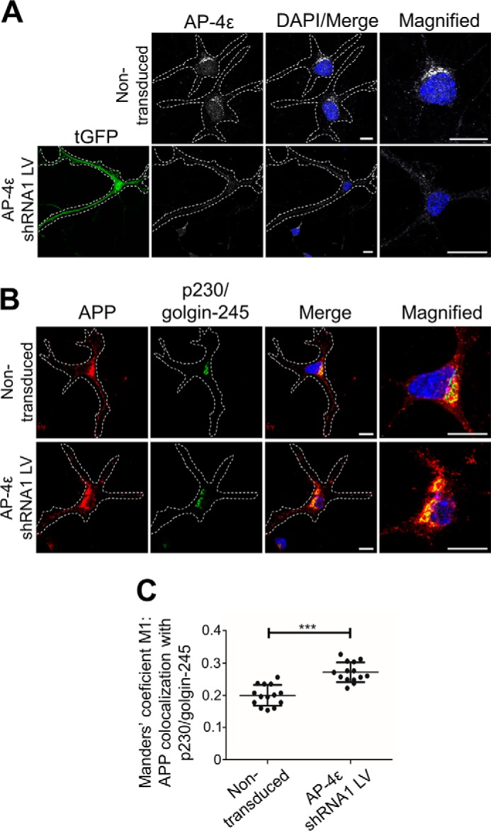
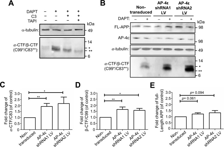
Amyloid precursor protein (APP) is processed along the amyloidogenic pathway by the β-secretase, BACE1, generating β-amyloid (Aβ), or along the nonamyloidogenic pathway by α-secretase, precluding Aβ production. The plasma membrane is considered the major site for α-secretase-mediated APP cleavage, but other cellular locations have not been rigorously investigated. Here, we report that APP is processed by endogenous α-secretase at the trans-Golgi network (TGN) of both transfected HeLa cells and mouse primary neurons. We have previously shown the adaptor protein complex, AP-4, and small G protein ADP-ribosylation factor-like GTPase 5b (Arl5b) are required for efficient post-Golgi transport of APP to endosomes. We found here that AP-4 or Arl5b depletion results in Golgi accumulation of APP and increased secretion of the soluble α-secretase cleavage product sAPPα. Moreover, inhibition of γ-secretase following APP accumulation in the TGN increases the levels of the membrane-bound C-terminal fragments of APP from both α-secretase cleavage (α-CTF, named C83 according to its band size) and BACE1 cleavage (β-CTF/C99). The level of C83 was ∼4 times higher than that of C99, indicating that α-secretase processing is the major pathway and that BACE1 processing is the minor pathway in the TGN. AP-4 silencing in mouse primary neurons also resulted in the accumulation of endogenous APP in the TGN and enhanced α-secretase processing. These findings identify the TGN as a major site for α-secretase processing in HeLa cells and primary neurons and indicate that both APP processing pathways can occur within the TGN compartment along the secretory pathway.

Keywords: adaptor protein; amyloid precursor protein (APP); enzyme processing; membrane trafficking; neurodegenerative disease; protein trafficking (Golgi); secretase; secretion; trans-Golgi network; α-secretase; β-secretase 1 (BACE1).


Conflict of interest statement

The authors declare that they have no conflicts of interest with the contents of this article

Figures

Figure 1.
Figure 1.
Detection of sAPPα in conditioned medium and CTFs in cell extracts. A, schematic of APP695 and epitope sequences of commercial antibodies. Monoclonal mouse anti-Aβ (clone W0-2; red) was raised against residues 4–10 of the Aβ domain. W0-2 recognizes full-length APP, sAPPα, Aβ peptide, and β-CTF/C99 fragments of APP. Monoclonal rabbit anti-APP (clone Y188; green) was raised against residues 682-687 (YENPTY motif) of APP695. Y188 recognizes full-length APP, β-CTF/C99, and α-CTF/C83 fragments of APP. B, HeLa cells stably expressing APP695WT (HeLa–APP695WT) were treated with either DMSO (carrier control) (−) or 250 nm γ-secretase inhibitor, DAPT (+), for 16 h. Conditioned medium (CM) and cell extracts (CE) were obtained. CM (10 μl) and CE (20 μg) were analyzed by immunoblotting with W0-2 or Y188 antibodies, using a chemiluminescence detection. The bands that correspond to sAPPα is marked (*). C, HeLa–APP695WT cell monolayers were treated either DMSO (carrier) or with 50 μm TAPI-1 (α-secretase inhibitor) for 6 h. Conditioned medium was collected, and equivalent volumes of samples, based on protein content of extracts of cell monolayers, were analyzed by immunoblotting with W0-2 antibodies. Cell extracts were analyzed by immunoblotting with mouse anti-α-tubulin antibodies. D, bar graph representing the percentage reduction of sAPPα levels in C. Data are represented as the mean ± S.D. of three independent experiments and analyzed by paired, two-tailed Student's t test. ***, p < 0.001. E, HeLa–APP695WT cell monolayers were treated either DMSO (carrier) or 250 nm DAPT in the presence or absence of 2 μm C3 (BACE1 inhibitor) and/or 5 μm TAPI-1 for 16–20 h. CM was analyzed by immunoblotting with W0-2 antibodies. CE were immunoblotted with either mouse anti-α-tubulin antibodies or rabbit anti-APP (Y188) antibodies. F, bar graph representing the percentage reduction of α-CTF/C83 levels in E. Data are represented as the mean ± S.D. of three independent experiments and analyzed by paired, two-tailed Student's t test. ***, p < 0.001.
Figure 2.
Figure 2.
AP-4ϵ depletion results in APP accumulation in the TGN. A, HeLa–APP695WT cells were transfected with either control siRNA, AP-4ϵ siRNA-1, or AP-4ϵ siRNA-2, as indicated, for 72 h. Monolayers were fixed and stained with mouse anti-human APP antibodies (clone NAB228) (red), rabbit anti-GCC88 antibodies (green) as a TGN marker, and DAPI (blue). Higher magnification of the merged images of the cells marked * are also shown. Bars represent 10 μm. B, percentage of APP at the TGN was calculated as a percentage of total APP pixels that overlapped with GCC88 using the OBCOL plugin on ImageJ. Data are represented as the mean ± S.D. of three independent experiments (n = 12) and analyzed by unpaired, two-tailed Student's t test. ***, p < 0.001.
Figure 3.
Figure 3.
Generation of sAPPα along the secretory pathway. A, HeLa–APP695WT cells or HeLa cells stably expressing constitutive active Arl5b(Q70L)-GFP were transfected with either control siRNA, AP-4ϵ siRNA, or Arl5b siRNA, as indicated, for 72 h and treated with either DMSO (carrier) or 250 nm DAPT in the final 16 h of siRNA transfection. Conditioned medium (10 μl) and cell extracts (20 μg) were subjected to SDS-PAGE and proteins transferred to a PVDF membrane. Conditional medium samples were probed with W0-2 antibodies and cell extract samples probed with either mouse anti-AP-4ϵ, mouse anti-AP1γ, mouse anti-GFP, or mouse anti-α-tubulin antibodies as indicated. B, bar graph representing fold change of sAPPα levels in AP-4ϵ siRNA or Arl5b siRNA-treated cells compared with control siRNA. Levels of sAPPα were normalized to total protein levels in cell extracts. Data are represented as the mean ± S.D. of three independent experiments and analyzed by paired, two-tailed Student's t test. ***, p < 0.001.
Figure 4.
Figure 4.
AP-4ϵ depletion results in accumulation of APP in cells. A, HeLa–APP695WT cells were transfected with either control siRNA or AP-4ϵ siRNA for 72 h and treated with either DMSO (carrier), 250 nm DAPT, 2 μm C3 (BACE1 inhibitor), or 250 nm DAPT + 2 μm C3 in the last 16 h of siRNA transfection. Cell extracts (20 μg) were analyzed by immunoblotting with either rabbit anti-APP (Y188), mouse anti-APP (W0-2), AP-4ϵ, or α-tubulin antibodies, as indicated. B, bar graph representing fold change of full-length APP levels in AP-4ϵ siRNA-treated cells compared with control siRNA. Levels of APP were normalized to α-tubulin. Data are represented as the mean ± S.D. of three independent experiments and analyzed by paired, two-tailed Student's t test. **, p < 0.01. C, quantitative PCR (qRT-PCR) analysis of human APP. Total RNA was purified from control siRNA and AP-4ϵ siRNA-treated cells, converted to cDNA, and singleplex reactions performed, in quadruplicate, for APP and the internal reference gene, GAPDH. qRT-PCR was performed and analyzed using the comparative ΔΔCT method. Relative quantification of mRNA levels used control siRNA cells as normalizer and GAPDH as the internal reference gene. Data are represented as the mean ± S.E. of two independent experiments.
Figure 5.
Figure 5.
Nonamyloidogenic processing of APP at the Golgi. A–D, quantitation of α-CTF/C83 levels from Fig. 4A. Shown are bar graphs representing fold change of α-CTF/C83 levels in AP-4ϵ siRNA–treated cells compared with control siRNA, under various treatment conditions as indicated. Levels of α-CTF/C83 were normalized to α-tubulin. Data are represented as the mean ± S.D. of three independent experiments and analyzed by paired two-tailed Student's t test. ***, p < 0.001.
Figure 6.
Figure 6.
Effect of α-secretase and γ-secretase inhibitors on secretion of the luminal domain of APP. A and B, HeLa–APP695WT cells were transfected with either control siRNA or AP-4ϵ siRNA for 72 h. Cells were also treated with either DMSO carrier (−), 250 nm DAPT (γ-secretase inhibitor), 2 μm C3 (BACE1 inhibitor), or 2 μm C3 and 250 nm DAPT (A) or 50 μm TAPI-1 (α-secretase inhibitor) or 50 μm TAPI-1 and 250 nm DAPT (B) in the last 16–20 h of siRNA transfection. Conditioned medium was collected, and the equivalent volume of samples based on protein content of extracts of cell monolayers were analyzed by immunoblotting with W0-2 antibodies or mouse anti-α-tubulin antibodies. CE were analyzed by immunoblotting with mouse anti-α-tubulin antibodies (the CM collected for analysis in A was carried out in conjunction with the experiment as Fig. 4A; therefore, the immunoblot for α-tubulin in the CE in Fig. 6A is the same as Fig. 4A).
Figure 7.
Figure 7.
Depletion of AP-4ϵ in mouse primary cortical neurons results in accumulation of APP in the TGN and increased APP processing. A and B, transduction of E16 primary mouse cortical neurons at DIV 3 with AP-4ϵ–shRNA lentivirus for 96 h. Neurons were fixed at DIV 7 and stained with either mouse anti-AP-4ϵ (gray) (A) or rabbit anti-APP (Y188; red) (B) and the TGN marker, human anti-p230/golgin-245 (far-red converted to green), antibodies overnight at 4 °C. Nuclei was stained with DAPI (blue). Bar represents 10 μm. C, VolocityTM software was used to calculate Manders' coefficient M1 values of APP colocalization with the TGN marker, p230/golgin-245. Data are represented as the mean ± S.D. of three independent experiments (n = 14) and analyzed by unpaired, two-tailed Student's t test. ***, p < 0.001.
Figure 8.
Figure 8.
Depletion of AP-4ϵ in mouse primary cortical neurons results in increased APP processing. A, E16 primary mouse cortical neurons were treated either DMSO (carrier) or 2 μm DAPT in the presence or absence of 2 μm C3 (BACE1 inhibitor) and/or 20 μm TAPI-1 for 16–20 h. Cell extracts were immunoblotted with either mouse anti-α-tubulin antibodies or rabbit anti-APP (Y188) antibodies to C99*/C83**. B, E16 primary mouse cortical neurons were transduced at DIV 3 with either AP-4ϵ shRNA1 or AP-4ϵ shRNA2 lentivirus (LV) for 96 h and treated with DMSO (carrier control) (−) or 2 μm DAPT in the last 16–20 h of transduction. Neurons were lysed in RIPA buffer and cell extracts (10 μg) subjected to SDS-PAGE, as described under “Experimental procedures.” Proteins transferred onto PVDF membrane were probed with mouse antibodies to either AP-4ϵ or α-tubulin. Proteins transferred onto nitrocellulose membrane were probed with rabbit anti-APP (Y188) antibodies to C99*/C83**. C–E, bar graph representing fold change of α-CTF/C83 (C), β-CTF/C99 (D), and full-length APP (E) levels in AP-4ϵ shRNA1 or AP-4ϵ shRNA2 lentiviral transduction compared with nontransduced controls. Levels of α-CTF/C83, β-CTF/C99, and full-length APP were normalized to α-tubulin. Data are represented as the mean ± S.D. of three independent experiments and analyzed by paired, two-tailed Student's t test. *, p < 0.05; **, p < 0.01.

References

    1. Thinakaran G., and Koo E. H. (2008) Amyloid precursor protein trafficking, processing, and function. J. Biol. Chem. 283, 29615–29619 10.1074/jbc.R800019200 - DOI - PMC - PubMed
    1. Kinoshita A., Fukumoto H., Shah T., Whelan C. M., Irizarry M. C., and Hyman B. T. (2003) Demonstration by FRET of BACE interaction with the amyloid precursor protein at the cell surface and in early endosomes. J. Cell Sci. 116, 3339–3346 10.1242/jcs.00643 - DOI - PubMed
    1. Koo E. H., Squazzo S. L., Selkoe D. J., and Koo C. H. (1996) Trafficking of cell-surface amyloid β-protein precursor. I. Secretion, endocytosis and recycling as detected by labeled monoclonal antibody. J. Cell Sci. 109, 991–998 - PubMed
    1. Rajendran L., Honsho M., Zahn T. R., Keller P., Geiger K. D., Verkade P., and Simons K. (2006) Alzheimer's disease β-amyloid peptides are released in association with exosomes. Proc. Natl. Acad. Sci. U.S.A. 103, 11172–11177 10.1073/pnas.0603838103 - DOI - PMC - PubMed
    1. Small S. A., and Gandy S. (2006) Sorting through the cell biology of Alzheimer's disease: intracellular pathways to pathogenesis. Neuron 52, 15–31 10.1016/j.neuron.2006.09.001 - DOI - PMC - PubMed

Publication types

Substances