Case Reports
. 2018 Dec 14:19:1488-1494.
doi: 10.12659/AJCR.912676.

Internal Hernia Associated with Colostomy After Laparoscopic Surgery for Rectal Malignancy: A Report of 3 Thought-Provoking Cases

Affiliations
Case Reports

Internal Hernia Associated with Colostomy After Laparoscopic Surgery for Rectal Malignancy: A Report of 3 Thought-Provoking Cases

Daiki Yasukawa et al. Am J Case Rep. .

Abstract

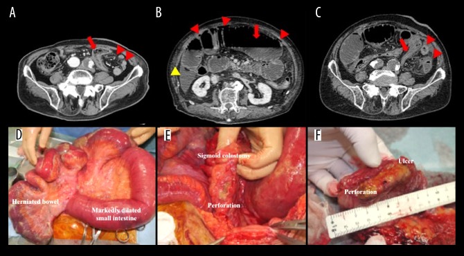
BACKGROUND Colostomy creation via intraperitoneal route is often performed during laparoscopic Hartmann's operation or abdominoperineal resection (APR). Herein, we report 3 rare cases of internal hernia associated with colostomy (IHAC). CASE REPORT The first case involved a 70-year-old man with IHAC after laparoscopic APR. Laparoscopy revealed the small intestine passed through a defect between the lifted sigmoid colon and left lateral abdominal wall in a cranial-to-caudal direction. The dislocated bowel with ischemic change was restored to its normal position and the lateral defect was covered with lateral peritoneum and greater omentum. The second case involved a 75-year-old man with IHAC after laparoscopic APR. Intraperitoneal findings were similar to those in the first case, except for the size of the lateral defect. This defect was too large for primary closure or patching; therefore, no surgical repair was performed. Unfortunately, this led to IHAC recurrence and creation of a new colostomy via extraperitoneal route. The third case involved an 85-year-old man with acute peritonitis resulting from IHAC after laparoscopic Hartmann's operation. Surgery revealed incarcerated bowels forming a closed loop and a perforation in the lifted sigmoid colon. The perforated colon was compressed by the dilated herniated bowel. The resected sigmoid colon showed perforation at the ulcer, which was shown on pathology to be caused by ischemia. CONCLUSIONS IHAC can lead not only to ischemia of strangulated bowel, but also to secondary damage to the lifted colon. During laparoscopic Hartmann's operation or APR, the colostomy should be created via extraperitoneal route to avoid IHAC.


Conflict of interest statement

Conflict of interest: None declared

Conflict of interest

None.

Figures

Figure 1.
Figure 1.
(A) Contrast-enhanced computed tomography shows part of the small intestine and its regional mesentery positioned in the lateral space between the lifted sigmoid colon and the lateral abdominal wall (arrowheads). The medially displaced and decompressed sigmoid colon is also observed (arrow). (B) The herniated small intestine in the pelvic space is dilated and fluid-filled but has persistent contrast uptake (arrowhead). A small amount of ascites is confirmed in the pelvis (arrow). (C) Laparoscopic exploration reveals the small intestine passing through the defect between the lifted sigmoid colon and left lateral abdominal wall in a cranial-to-caudal direction. The incarcerated bowel forms a closed loop with ischemic changes. (D) The lateral defect was covered with lateral peritoneum and greater omentum to avoid postoperative recurrence of internal hernia associated with colostomy.
Figure 2.
Figure 2.
(A) Contrast-enhanced computed tomography shows part of the small intestine and its regional mesentery positioned in the lateral space between the lifted sigmoid colon and lateral abdominal wall (arrowheads). The medially displaced and decompressed sigmoid colon is observed (arrow). (B) The herniated small intestine in the pelvic space is dilated and fluid-filled but has persistent contrast uptake (arrowhead). (C, D) Laparoscopic exploration revealed that the small intestine passed through the defect between the lifted sigmoid colon and left lateral abdominal wall in a cranial-to-caudal direction, and that the incarcerated bowel formed a closed loop. This malposition resulted in partial occlusion of blood flow.
Figure 3.
Figure 3.
(A) Follow-up contrast-enhanced computed tomography 2 months after surgery shows the small intestine passing through a defect between the lifted sigmoid colon and left lateral abdominal wall (arrowheads) in a cranial-to-caudal direction. No disturbance of bowel passage was observed, and the lifted sigmoid colon is seen (arrow). (B) Contrast-enhanced computed tomography 2 days after hospitalization reveals intraperitoneal free air (red arrowheads) and marked dilation of the small intestine (arrow) with ascites (yellow arrowhead). (C) Contrast-enhanced computed tomography shows part of the small intestine and regional mesentery positioned in the lateral space between the lifted sigmoid colon and lateral abdominal wall (arrowheads). The medially displaced and decompressed sigmoid colon is observed (arrow). (D) Emergent laparotomy revealed that the small intestine passed through the defect between the lifted sigmoid colon and left lateral abdominal wall in a cranial-to-caudal direction. The incarcerated bowel formed a closed loop and had ischemic color change. (E) A perforation was identified in the lifted sigmoid colon, which was secondarily compressed by the dilated herniated bowel. (F) Resected specimen had a perforation at the site of ulceration, which was shown on pathology to be caused by ischemia.
Figure 4.
Figure 4.
Colostomy is created, via an extraperitoneal route to prevent IHAC (red arrow) even in fewer adhesions by laparoscopic surgery, and via a staggered fascia incisions (arrowheads) to avoid common parastomal hernia (blue arrow).

References

    1. Scala D, Niglio A, Pace U, et al. Laparoscopic intersphincteric resection: Indications and results. Updates Surg. 2016;68:85–91. - PubMed
    1. Mariani P, Ghanneme A, De la Rochefordiere A, et al. Abdominoperineal resection for anal cancer. Dis Colon Rectum. 2008;51:1495–501. - PubMed
    1. Robertson I, Leung E, Hughes D, et al. Prospective analysis of stoma-related complications. Colorectal Dis. 2005;7:279–85. - PubMed
    1. Xu Z, Guo W. [Clinical research progress of mesenteric internal hernia after Roux-en-Y reconstruction] Zhonghua Wei Chang Wai Ke Za Zhi. 2017;20:352–56. [in Chinese] - PubMed
    1. Toh JW, Lim R, Keshava A, Rickard MJ. The risk of internal hernia or volvulus after laparoscopic colorectal surgery: A systematic review. Colorectal Dis. 2016;18:1133–41. - PubMed

Publication types