. 2018 Dec 13;9(1):5301.
doi: 10.1038/s41467-018-07579-y.

Tissue engineering of human hair follicles using a biomimetic developmental approach

Affiliations

Tissue engineering of human hair follicles using a biomimetic developmental approach

Hasan Erbil Abaci et al. Nat Commun. .

Abstract

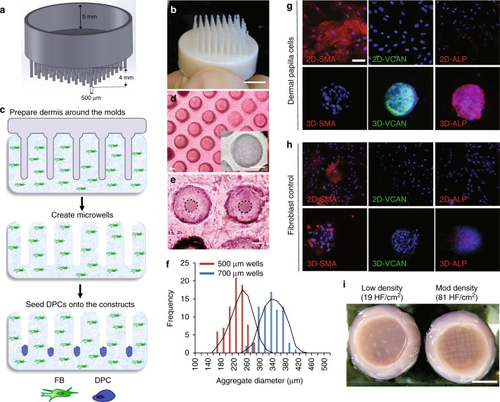
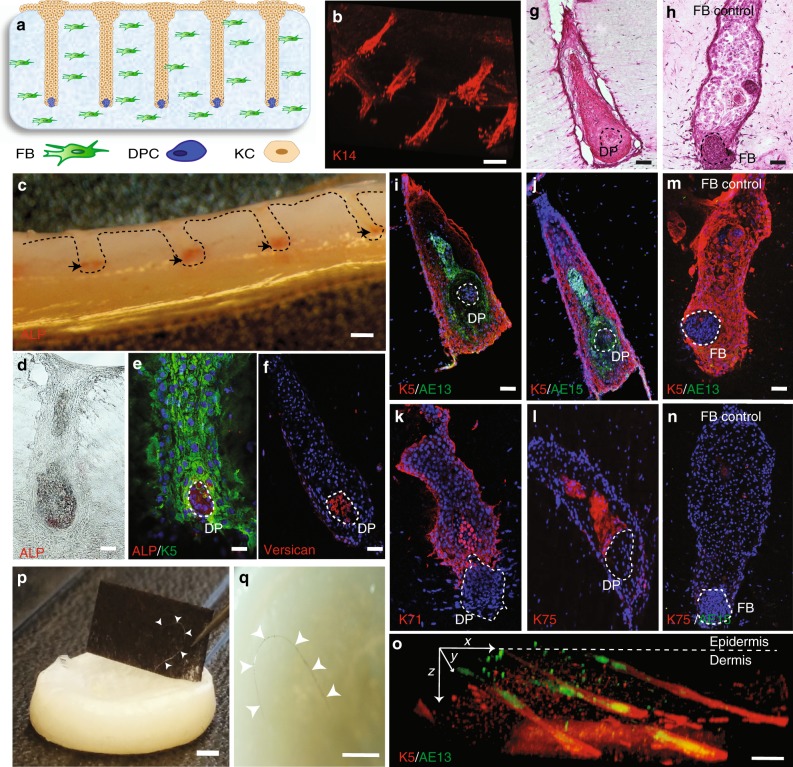
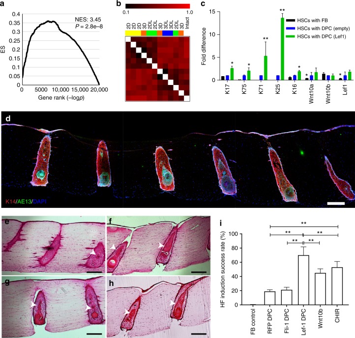
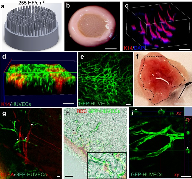
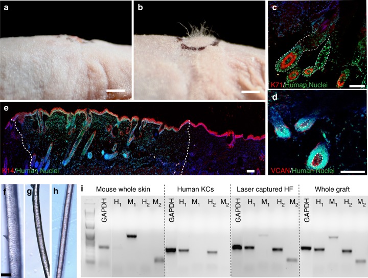
Human skin constructs (HSCs) have the potential to provide an effective therapy for patients with significant skin injuries and to enable human-relevant drug screening for skin diseases; however, the incorporation of engineered skin appendages, such as hair follicles (HFs), into HSCs remains a major challenge. Here, we demonstrate a biomimetic approach for generation of human HFs within HSCs by recapitulating the physiological 3D organization of cells in the HF microenvironment using 3D-printed molds. Overexpression of Lef-1 in dermal papilla cells (DPC) restores the intact DPC transcriptional signature and significantly enhances the efficiency of HF differentiation in HSCs. Furthermore, vascularization of hair-bearing HSCs prior to engraftment allows for efficient human hair growth in immunodeficient mice. The ability to regenerate an entire HF from cultured human cells will have a transformative impact on the medical management of different types of alopecia, as well as chronic wounds, which represent major unmet medical needs.


Conflict of interest statement

A.M.C. and C.A.J. are founders of Rapunzel Bioscience Inc, focused on developing regenerative therapies for skin and hair disorders. The remaining authors declare no competing interests.

Figures

Fig. 1
Fig. 1
Patterning of collagen type-1 gel using 3D-printed molds allows for physiological arrangement of cells in the hair follicle. Hair follicle molds were designed (a) and 3D-printed (b) to have HF-like extensions and a 5-mm-deep cavity that allows the molds to float on collagen gel. c The collagen gel containing dermal fibroblasts was allowed to solidify around the HF-like extensions to create an array of microwells in which the DPCs formed spontaneous aggregates. d Top view of HSCs containing DPCs which settled down into the microwells (lower panel: higher magnification of the microwells). e DPCs formed spontaneous aggregates at the center of the microwells (dashed line circles the aggregates). f The size of DPC aggregates, as calculated from images, was correlated with the diameter of the microwells (500 µm wells in red and 700 µm wells in blue) (n = 9; three technical and three biological replicates). Expression of SMA and VCAN and the activity of ALP in 2D cultures of DPCs and in DPCs aggregates formed in the microwells (g) was compared to 2D cultures of FBs and FB aggregates in the microwells (h). i Top view of the 3D-reconstructed dermis at two different hair follicle densities of 19 HF per cm2 and 81 HF per cm2. Scale bars for (d) and (eh) are 100 µm and 5 mm for (b) and (i)
Fig. 2
Fig. 2
Differentiation of human keratinocytes into specific hair follicle lineages in HSCs. a Schematic and b 3D-reconstructed image of K14-positive cells showing microwells filled with KCs after a day of culture. c Cross section of the whole HSCs with the ALP-active labeled cells at the tip of the microwells (black arrows). Bright-field (d) and e, f immunofluorescent images of HSCs reveal the physiological conformation of cells where ALP-active and VCAN expressing DPCs (dashed circles) are engulfed by K5-positive KCs after a day of culture, forming HFUs. H&E staining of HSCs depicted the morphological changes of KCs above DPC aggregates (g), which was not observed in HSCs with FB aggregates (h). KCs in the HSCs expressed specific hair lineage and differentiation markers K5 and AE13 (i), AE15 (j), K71 (k), and K75 (l), which was not observed in HSCs with FB aggregates (m, n) after a week of culture. o Culture of HSCs for 3 weeks resulted in elongated hair follicles and improved organization of inner and outer root sheaths as shown by the K5 and AE13 markers, respectively. p, q Prolonged culture period led to hair fiber formation and protrusion from the HSCs (arrow heads indicate the hair fiber). Scale bars for (b), (c) and (oq) are 2 mm and for (dn) are 100 µm (n = 9; three technical and three biological replicates)
Fig. 3
Fig. 3
Overexpression of the master regulator gene Lef-1 in DPCs enhances the efficiency of hair follicle induction in HSCs. a GSEA of the RNA-sequencing data of Lef-1-transfected DPCs revealed that Lef-1 overexpression significantly overlaps the downstream network genes previously predicted by ARACNE algorithm (NES, normalized enrichment score). b Gene distance matrix showing the individual and synergistic effects of Lef-1 overexpression and 3D-spheroid culture in DPC transcriptional signature (2D: adherent culture; 3D: spheroid culture; 2DL: adherent culture of Lef-1-transfected cells; 3DL: spheroid culture of Lef-1-transfected cells). c Expression levels of specific hair follicle layers and Wnt-signaling genes determined by qPCR from the total mRNA of HSCs containing Lef-1-transfected DPCs (green), empty vector-transfected DPCs (blue) and FBs control (black). Fold difference values are based on the empty vector-transfected DPCs. d Low magnification image demonstrating expression of AE13 in HSCs generated using Lef-1-transfected DPCs and the high efficiency in HF lineage differentiation. Histological H&E staining of the constructs generated by empty vector-transfected DPCs (e), Lef-1-transfected DPCs (f), Wnt10b-treated DPCs (g), and CHIR99021-treated DPCs (h) (arrows showing differentiated KC morphology). i Success rate, defined as the ratio of the HFUs exhibiting hair follicle differentiation to the total number of HFUs, for HSCs with DPCs at different treatment conditions compared to FB control (n = 3 with cells from three donors, *p < 0.05 and **p < 0.005 from two-tailed t test); Center values and error bars are defined as means and s.e.m.; Scale bars are 300 µm
Fig. 4
Fig. 4
Vascularization of high hair-follicle-density HSCs for efficient engraftment. a, b HSCs were generated using the molds that have 255 HF per cm2 to promote hair growth in grafts. c 3D-reconstructed wholemount image of the HSCs showing 3D conformation of K14-positive cells at the high hair follicle density. GFP-tagged HUVECs that were encapsulated in the dermal compartment with the fibroblasts closely surrounded the K14-positive cells in the hair follicle structures (d) and formed capillary-like networks after 3 days of culture (e). f Bottom view of the explanted prevascularized HSCs (dashed circle) with GFP-HUVECs showing promoted host vascularization and blood supply to the grafts (n = 9; three technical and three biological replicates); g HSCs grafted onto mice were revascularized by the host vessels where GFP-tagged HUVECs (green) formed capillaries that are well-organized around the mouse vessels (GS-IB4 staining in red; arrow heads show the proximity of mouse and human vessels) and red blood cells (RBCs) (h), (arrow heads show host RBCs near human blood vessels and lower panel shows a magnified area of the image); and exhibited lumen formation (arrow heads show the lumen) (i). Scale bars for (b and f), (c, d), and (e, g, h and i) are 4 mm, 2 mm, and 100 µm, respectively
Fig. 5
Fig. 5
Induction of human hair growth in immune-deficient nude mice. a, b Engraftment of high follicle-density HSCs onto immune-deficient nude mice led to hair growth in the grafts after 4–6 weeks (b), which was not observed in the HSCs prepared with FB aggregates as a control (a) (n = 10; five biological and two technical replicates per each condition). c, d Human-specific nuclear staining (green) indicated that the de novo hair follicles marked with K71 (c) (HF boundaries circled in dashed line) and DPCs marked with VCAN (d) are comprised of human cells (DPs circled in dashed line). e Low magnification image of the explanted grafts revealed the boundaries (dashed line) between the mouse and human tissues as marked by the human nuclear staining. Bright-field microscopy of unpigmented terminal human hair (f), engineered human hair in the grafts (g), and unpigmented human vellus hair (h) showed morphological similarities between human hair and engineered hair. i PCR was performed with two sets of primers specific to human and mouse cells using the RNA of the laser captured hair follicles from the grafts in comparison to whole grafts, mouse skin, and cultured human KCs. Scale bars for (a, b), (ce) and (fh) are 2 mm, 200 µm, and 50 µm respectively

References

    1. Sen CK, et al. Human skin wounds: a major and snowballing threat to public health and the economy. Wound Repair Regen. 2009;17:763–771. doi: 10.1111/j.1524-475X.2009.00543.x. - DOI - PMC - PubMed
    1. Boyce ST, et al. Cultured skin substitutes reduce requirements for harvesting of skin autograft for closure of excised, full-thickness burns. J. Trauma. 2006;60:821–829. - PubMed
    1. Abaci HE, Gledhill K, Guo Z, Christiano AM, Shuler ML. Pumpless microfluidic platform for drug testing on human skin equivalents. Lab Chip. 2015;15:882–888. doi: 10.1039/C4LC00999A. - DOI - PMC - PubMed
    1. Schneider MR, Schmidt-Ullrich R, Paus R. The hair follicle as a dynamic miniorgan. Curr. Biol. 2009;19:R132–R142. doi: 10.1016/j.cub.2008.12.005. - DOI - PubMed
    1. Abaci HE, et al. Human skin constructs with spatially controlled vasculature using primary and iPSC-derived endothelial cells. Adv. Healthc. Mater. 2016;5:1800–1807. doi: 10.1002/adhm.201500936. - DOI - PMC - PubMed

Publication types

Substances