. 2018 Dec 13;9(1):5308.
doi: 10.1038/s41467-018-07775-w.

Virus-mediated export of chromosomal DNA in plants

Affiliations

Virus-mediated export of chromosomal DNA in plants

Marco Catoni et al. Nat Commun. .

Abstract

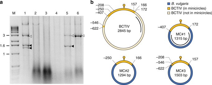
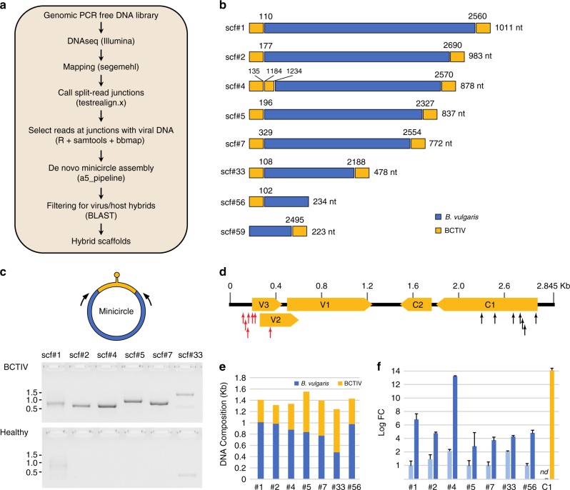
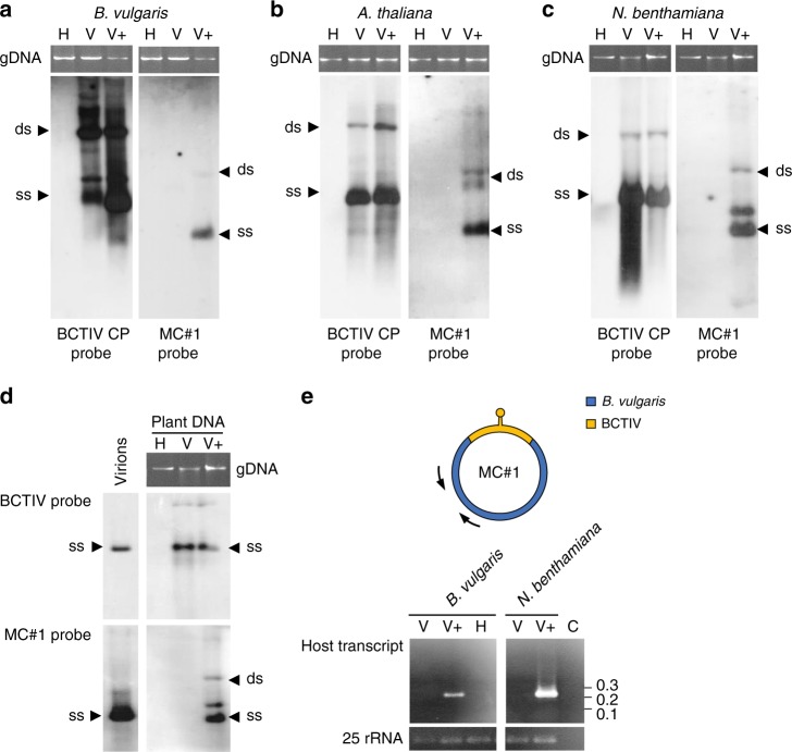
The propensity of viruses to acquire genetic material from relatives and possibly from infected hosts makes them excellent candidates as vectors for horizontal gene transfer. However, virus-mediated acquisition of host genetic material, as deduced from historical events, appears to be rare. Here, we report spontaneous and surprisingly efficient generation of hybrid virus/host DNA molecules in the form of minicircles during infection of Beta vulgaris by Beet curly top Iran virus (BCTIV), a single-stranded DNA virus. The hybrid minicircles replicate, become encapsidated into viral particles, and spread systemically throughout infected plants in parallel with the viral infection. Importantly, when co-infected with BCTIV, B. vulgaris DNA captured in minicircles replicates and is transcribed in other plant species that are sensitive to BCTIV infection. Thus, we have likely documented in real time the initial steps of a possible path of virus-mediated horizontal transfer of chromosomal DNA between plant species.


Conflict of interest statement

The authors declare no competing interests.

Figures

Fig. 1
Fig. 1
Identification of chimeric minicircles in field-grown sugar beet plants. a DNA fragments obtained after RCA and EcoRI digestion of DNA samples from different plants (lanes 1–6) collected from a field in Iran. The putative full-length BCTIV genome and smaller molecules are indicated by white and black arrows, respectively. Lane M, molecular markers with the size (kb) indicated on the left. Source data are provided as Supplementary Data 5. b Schematic representation of BCTIV and the three minicircles obtained from some of the BCTIV-infected field samples shown in a; MC#1 originates from plant 5, MC#2 and #3 from plant 1. The BCTIV and the minicircle genomes are represented in circular form; yellow and blue represent the BCTIV and the B. vulgaris genome-derived elements, respectively. The recombination coordinates on the BCTIV genome and the minicircles are indicated in nucleotides in relation to the conventional start of the BCTIV genome located at the stem-loop region (marked as a yellow lollipop). The locations of the MC#1- and BCTIV-specific probes used for subsequent southern blot analysis are represented by black lines. The complete minicircle sequences are reported in Supplementary Data 2
Fig. 2
Fig. 2
Identification of minicircles in experimentally infected Beta vulgaris plants by the NGS approach. a Diagram of the bioinformatic pipeline used for the detection of minicircles. The tools used in each step are indicated in parentheses (refer to the Methods section for details). b Schematic representation of the eight BCTIV/Beta vulgaris hybrid scaffolds assembled after the filtering step described in a. The junction coordinates referring to the BCTIV genome are indicated above each recombination point. The sizes (nt) of the B. vulgaris DNA fragments (blue portions) are reported on the right. c In vivo validation of the circular nature of the scaffolds reported in b, performed by inverse PCR (see also Supplementary Figure 3). The approximate positions of the primers specific for the non-viral portions of each minicircle (in blue) are indicated by arrows. DNA from mock-inoculated B. vulgaris was used as a negative control. Molecular markers (kb) are shown on the side. Source data are provided as Supplementary Data 5. d Distribution of minicircle junctions between viral and host DNA. The proximal and distal junctions are marked with red and black arrows, respectively. BCTIV ORFs are represented by yellow boxes. e Relative contributions of B. vulgaris and BCTIV DNA to minicircles identified by NGS. f Relative increase in copy number of chromosomal DNA fragments due to their amplification as parts of minicircles in the BCTIV-infected plant. Copy number was determined by quantitative PCR and normalized to a single copy gene (GAPDH). Light-blue bars represent healthy plants and dark-blue bars BCTIV-infected plants. BCTIV copy number was estimated by amplification of a fragment of the C1 ORF (yellow bar). Each bar represents the mean of three repetitions; ±s.d. marked by error bars; nd not detected. Source data are provided as Supplementary Data 5
Fig. 3
Fig. 3
MC#1 replication and transcription when transfected to various hosts. Southern blots of DNA extracted from B. vulgaris (a), A. thaliana (b) and N. benthamiana (c), mock-inoculated healthy plants (H); plants agroinfected with BCTIV (V) or with both BCTIV and MC#1 (V+). Leaves were collected at 4 weeks post inoculation (wpi) and hybridized with BCTIV- or MC#1-specific probes (see Fig. 1b). Bands of single and double stranded DNAs are indicated as “ss” and “ds”, respectively. Plant genomic DNA (gDNA) is shown above the blots as a loading control. Source data are provided as Supplementary Data 5. d Southern blot of DNA extracted from purified BCTIV virions and from the N. benthamiana plants used for purification of BCTIV particles, hybridized with the BCTIV- and the MC#1-specific probes (see Fig. 1b). “ss”, “ds” and gDNA as in ac. Source data are provided as Supplementary Data 5. e Transcription of the beet-derived DNA sequence of MC#1 in B. vulgaris and N. benthamiana plants inoculated with BCTIV and MC#1 (V+) detected by RT-PCR with primers positioned as indicated by arrows in the upper part of the panel. Lanes are marked H, V and V+ as in ad, whereas C is a RT-PCR-negative reaction control. Source data are provided as Supplementary Data 5

References

    1. Keeling PJ, Palmer JD. Horizontal gene transfer in eukaryotic evolution. Nat. Rev. Genet. 2008;9:605–618. doi: 10.1038/nrg2386. - DOI - PubMed
    1. El Baidouri M, et al. Widespread and frequent horizontal transfers of transposable elements in plants. Genome Res. 2014;24:831–838. doi: 10.1101/gr.164400.113. - DOI - PMC - PubMed
    1. Schaack S, Gilbert C, Feschotte C. Promiscuous DNA: horizontal transfer of transposable elements and why it matters for eukaryotic evolution. Trends Ecol. Evol. 2010;25:537–546. doi: 10.1016/j.tree.2010.06.001. - DOI - PMC - PubMed
    1. Gilbert C, Cordaux R. Viruses as vectors of horizontal transfer of genetic material in eukaryotes. Curr. Opin. Virol. 2017;25:16–22. doi: 10.1016/j.coviro.2017.06.005. - DOI - PubMed
    1. Ekblom R, Galindo J. Applications of next generation sequencing in molecular ecology of non-model organisms. Heredity. 2011;107:1–15. doi: 10.1038/hdy.2010.152. - DOI - PMC - PubMed

Publication types

Supplementary concepts