. 2018 Dec 13;9(1):5300.
doi: 10.1038/s41467-018-07747-0.

Vagus-macrophage-hepatocyte link promotes post-injury liver regeneration and whole-body survival through hepatic FoxM1 activation

Affiliations

Vagus-macrophage-hepatocyte link promotes post-injury liver regeneration and whole-body survival through hepatic FoxM1 activation

Tomohito Izumi et al. Nat Commun. .

Abstract

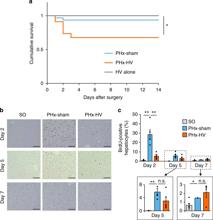
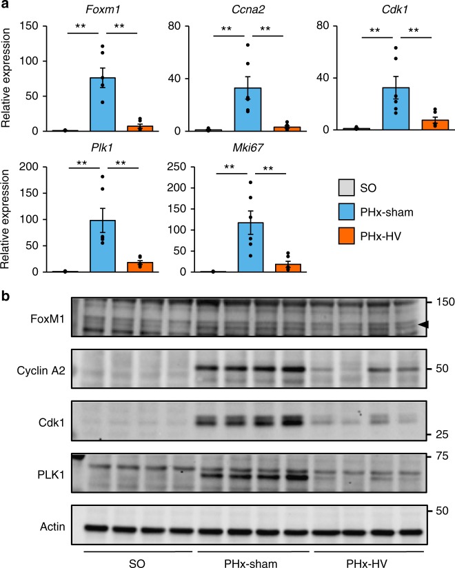
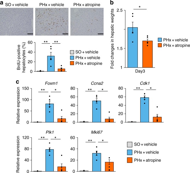
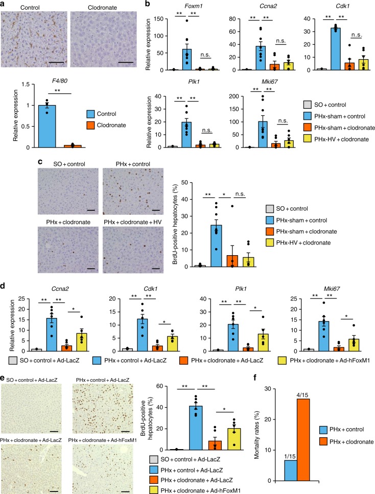
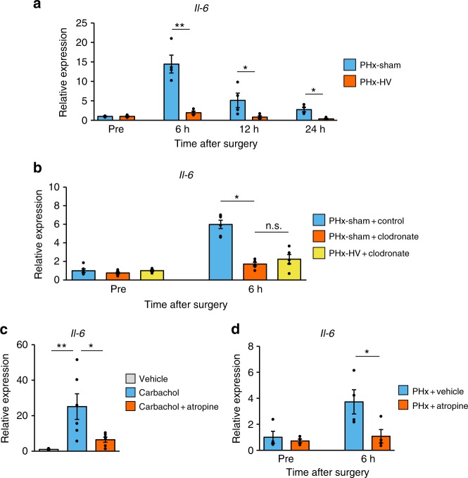
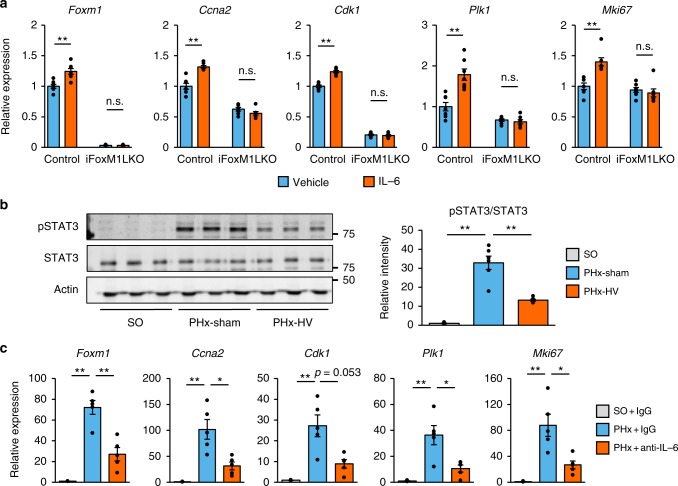
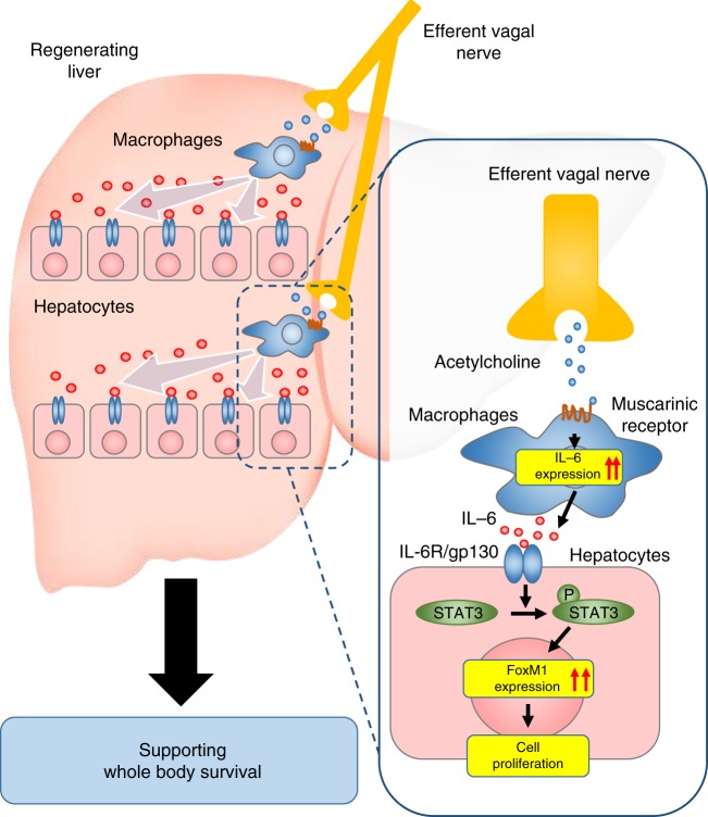
The liver possesses a high regenerative capacity. Liver regeneration is a compensatory response overcoming disturbances of whole-body homeostasis provoked by organ defects. Here we show that a vagus-macrophage-hepatocyte link regulates acute liver regeneration after liver injury and that this system is critical for promoting survival. Hepatic Foxm1 is rapidly upregulated after partial hepatectomy (PHx). Hepatic branch vagotomy (HV) suppresses this upregulation and hepatocyte proliferation, thereby increasing mortality. In addition, hepatic FoxM1 supplementation in vagotomized mice reverses the suppression of liver regeneration and blocks the increase in post-PHx mortality. Hepatic macrophage depletion suppresses both post-PHx Foxm1 upregulation and remnant liver regeneration, and increases mortality. Hepatic Il-6 rises rapidly after PHx and this is suppressed by HV, muscarinic blockade or resident macrophage depletion. Furthermore, IL-6 neutralization suppresses post-PHx Foxm1 upregulation and remnant liver regeneration. Collectively, vagal signal-mediated IL-6 production in hepatic macrophages upregulates hepatocyte FoxM1, leading to liver regeneration and assures survival.


Conflict of interest statement

The authors declare no competing interests.

Figures

Fig. 1
Fig. 1
Vagal signals are critical for post PHx survival. a Cumulative survival of mice that underwent PHx-sham (n = 26), PHx-HV (n = 26), and HV alone (n = 18). b Representative images of liver sections immunostained for BrdU on days 2, 5, and 7 after sham operation for PHx (SO), PHx-sham, or PHx-HV. Scale bars indicate 100 µm. c BrdU-positive hepatocyte ratios in SO mice (n = 5–6 per group), PHx-sham mice (n = 4–6 per group), and PHx-HV mice (n = 4–6 per group) on postoperative days 2, 5, and 7. The magnified graphs on days 5 and 7 are shown in framed boxes with lowered scale ranges. *P< 0.05; **P < 0.01 assessed by log-rank test with Bonferroni correction (a) or one-way ANOVA followed by Bonferroni’s post hoc test (c). n.s., not significant
Fig. 2
Fig. 2
Vagal signals are involved in activation of hepatic FoxM1 after PHx. a Relative expressions of Foxm1, its target genes. and MKi67 in the liver 2 days after SO (n = 4), PHx-sham (n = 6), and PHx-HV (n = 6). b Images of liver extract immunoblottings with anti-FoxM1, Cyclin A2, Cdk1, PLK1, and actin in SO, PHx-sham, and PHx-HV mice on postoperative day 2. Arrowhead indicates bands for FoxM1. Data are presented as means ± SEM. *P < 0.01 assessed by one-way ANOVA followed by Bonferroni’s post hoc test (a). n.s., not significant
Fig. 3
Fig. 3
FoxM1 activation promotes liver regeneration and whole-body survival. a Relative expressions of Foxm1, its target genes, and MKi67 in the liver 2 days after surgery in control mice that underwent SO (n = 5) and PHx (n = 5), and iFoxM1LKO mice that underwent PHx (n = 5). b (Upper panels) Representative images of liver sections immunostained for BrdU 2 days after PHx from control and iFoxM1LKO mice. Scale bars indicate 100 µm. (Lower panel) BrdU-positive hepatocyte ratios in control (n = 5) and iFoxM1LKO (n = 5) mice 2 days after PHx. c Relative expressions of Foxm1, its target genes, and MKi67 in the liver 2 days after SO (n = 5), PHx-sham (n = 6), and PHx-HV (n = 5) in mice receiving Ad-LacZ and after PHx-HV in mice receiving Ad-hFoxM1 (n = 6). d (Upper panels) Representative images of liver sections immunostained for BrdU 2 days after SO, PHx-sham, and PHx-HV from mice receiving Ad-LacZ and after PHx-HV from mice receiving Ad-hFoxM1. Scale bars indicate 100 µm. (Lower panel) BrdU-positive hepatocyte ratios in the liver 2 days after SO (n = 5), PHx-sham (n = 6), and PHx-HV (n = 5) in Ad-LacZ-treated mice and PHx-HV in Ad-hFoxM1-treated mice (n = 6). e Cumulative survival of mice that underwent PHx-sham (n = 21) and PHx-HV (n = 34) procedures after receiving Ad-LacZ and mice that underwent PHx-HV after receiving Ad-hFoxM1 (n = 34). Data are presented as means ± SEM. *P< 0.05; **P < 0.01 assessed by one-way ANOVA followed by Bonferroni’s post hoc test (a, c, d), unpaired t test (b), or log-rank test with Bonferroni correction (e). n.s., not significant
Fig. 4
Fig. 4
Muscarinic signals are involved in acute liver regenerative responses after PHx. a (Upper panels) Representative images of liver sections immunostained for BrdU on day 2 after sham operation for PHx (SO) and PHx in vehicle-treated mice and after PHx in atropine-treated mice. Scale bars indicate 100 µm. (Lower panel) BrdU-positive hepatocyte ratios in SO + vehicle mice (n = 4), PHx + vehicle mice (n = 5), and PHx + atropine mice (n = 5). b Fold changes in hepatic weights 3 days after surgery in PHx + vehicle mice (n = 5) and PHx + atropine mice (n = 5). Hepatic weights were divided by those obtained immediately after surgery. c Relative expressions of Foxm1, its target genes, and MKi67 in the liver 2 days after SO (n = 4) and PHx (n = 5) in vehicle-treated mice and PHx (n = 5) in atropine-treated mice. *P< 0.05; **P < 0.01 assessed by one-way ANOVA followed by Bonferroni’s post hoc test (a and c) or assessed by unpaired t test (b). n.s., not significant
Fig. 5
Fig. 5
Macrophages mediate vagus-induced hepatic FoxM1 activation. a (Upper panels) Representative images of liver sections immunostained for F4/80 from mice 24 h after receiving control and clodronate liposomes. Scale bars indicate 100 µm. (Lower panel) Relative gene expressions of F4/80 in the liver 24 h after control (n = 4) and clodronate (n = 4) liposome administration. b Relative expressions of Foxm1, its target genes, and MKi67 in the liver 2 days after surgery in SO + control mice (n = 6), PHx-sham + control mice (n = 8), PHx-sham + clodronate mice (n = 6), and PHx-HV + clodronate mice (n = 6). c (Left panels) Representative images of liver sections immunostained for BrdU on day 2 in SO + control-, PHx-sham + control-, PHx-sham + clordronate-, and PHx-HV + clodronate mice. Scale bars indicate 100 µm. (Right panel) BrdU-positive hepatocyte ratios in SO + control mice (n = 6), PHx-sham + control mice (n = 8), PHx-sham + clodronate mice (n = 6,) and PHx-HV + clodronate mice (n = 6) 2 days after surgery. d Relative expressions of Foxm1, its target genes, and MKi67 in the liver 2 days after SO in mice receiving control liposome and Ad-LacZ (n = 6) and PHx in mice receiving control liposome and Ad-LacZ (n = 6), clodronate liposome and Ad-LacZ (n = 6), and clodronate liposome and Ad-hFoxM1 (n = 6). e (Left panels) Representative images of liver sections immunostained for BrdU on day 2 after SO in mice receiving control liposome and Ad-LacZ and PHx in mice receiving control liposome and Ad-LacZ, clodronate liposome and Ad-LacZ, and clodronate liposome and Ad-hFoxM1. Scale bars indicate 100 µm. (Right panel) BrdU-positive hepatocyte ratios in the liver 2 days after SO in mice receiving control liposome and Ad-LacZ (n = 6) and PHx in mice receiving control liposome and Ad-LacZ (n = 6), clodronate liposome and Ad-LacZ (n = 6), and clodronate liposome and Ad-hFoxM1 (n = 6). f Mortality rates within 2 postoperative days in PHx + control mice (n = 15) and PHx + clodronate mice (n = 15). Data are presented as means ± SEM. *P< 0.05; **P < 0.01 assessed by one-way ANOVA followed by Bonferroni’s post hoc test (b, c, d, e) or assessed by unpaired t test (a). n.s., not significant
Fig. 6
Fig. 6
Vagal signals increase macrophage IL-6 production after PHx. a Relative gene expressions of Il-6 after surgery in PHx-sham mice (n = 4 per group) and PHx-HV mice (n = 4 per group). b Relative gene expressions of Il-6 after PHx-sham in control liposome-treated (n = 5–6 per group) and clodronate liposome-treated mice (n = 5–6 per group), and after PHx-HV in clodronate liposome-treated mice (n = 5–6 per group). c Relative gene expression of Il-6 in primary macrophages after 4 h of stimulation with vehicle (n = 6), 100 µM carbachol (n = 6), or both 100 µM carbachol and 100 µM atropine (n = 6). d Relative gene expressions of Il-6 after PHx in vehicle-treated (n = 4 per group) and atropine-treated (n = 4 per group) mice. *P< 0.05; **P < 0.01 assessed by unpaired t test (a and d), or assessed by one-way ANOVA followed by Bonferroni’s post hoc test (b and c). n.s., not significant
Fig. 7
Fig. 7
IL-6 mediates vagal signal-induced hepatic FoxM1 activation. a Relative expressions of Foxm1, its target genes, and MKi67 in primary hepatocytes isolated from control and iFoxM1LKO mice treated with 100 ng/ml IL-6 (n = 7–8 per group) and vehicle (n = 7–8 per group) for 6 h. b (Left panels) Representative images of liver extract immunoblottings with anti-phospho-STAT3, total STAT3, and actin. (Right panel) Relative intensities of phospho/total STAT3 in livers from sham operation for PHx (SO)- (n = 6), PHx-sham- (n = 6), and PHx-HV mice (n = 6). c Relative expressions of Foxm1, its target genes, and MKi67 in the liver 2 days after surgery from SO + IgG (n = 5), PHx + IgG (n = 5), and PHx + anti-IL-6 antibody- (n = 5) treated mice. Data are presented as means ± SEM. *P< 0.05; **P < 0.01 assessed by unpaired t test (a), or assessed by one-way ANOVA followed by Bonferroni’s post hoc test (b and c). n.s., not significant
Fig. 8
Fig. 8
The proposed mechanism of vagus-mediated liver regeneration. The multistep regulatory mechanism of acute liver regeneration after PHx via a vagus–macrophage–hepatocyte link

Similar articles

Cited by

References

    1. Michalopoulos GK, DeFrances MC. Liver regeneration. Science. 1997;276:60–66. doi: 10.1126/science.276.5309.60. - DOI - PubMed
    1. Michalopoulos GK. Hepatostat: liver regeneration and normal liver tissue maintenance. Hepatology. 2017;65:1384–1392. doi: 10.1002/hep.28988. - DOI - PubMed
    1. Ikeda O, et al. Autonomic regulation of liver regeneration after partial hepatectomy in mice. J. Surg. Res. 2009;152:218–223. doi: 10.1016/j.jss.2008.02.059. - DOI - PubMed
    1. Kato H, Shimazu T. Effect of autonomic denervation on DNA synthesis during liver regeneration after partial hepatectomy. Eur. J. Biochem. 1983;134:473–478. doi: 10.1111/j.1432-1033.1983.tb07591.x. - DOI - PubMed
    1. Ohtake M, Sakaguchi T, Yoshida K, Muto T. Hepatic branch vagotomy can suppress liver regeneration in partially hepatectomized rats. HPB Surg. 1993;6:277–286. doi: 10.1155/1993/59691. - DOI - PMC - PubMed

Publication types

LinkOut - more resources