. 2018 Dec 13;9(1):5305.
doi: 10.1038/s41467-018-07753-2.

Intron-containing RNA from the HIV-1 provirus activates type I interferon and inflammatory cytokines

Affiliations

Intron-containing RNA from the HIV-1 provirus activates type I interferon and inflammatory cytokines

Sean Matthew McCauley et al. Nat Commun. .

Abstract

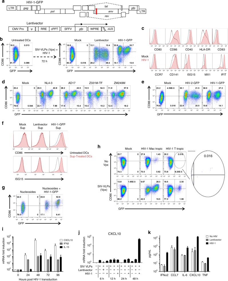
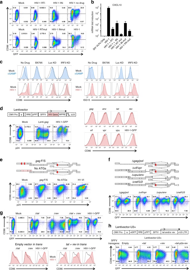
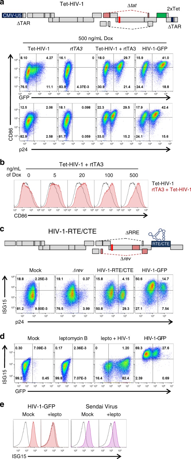
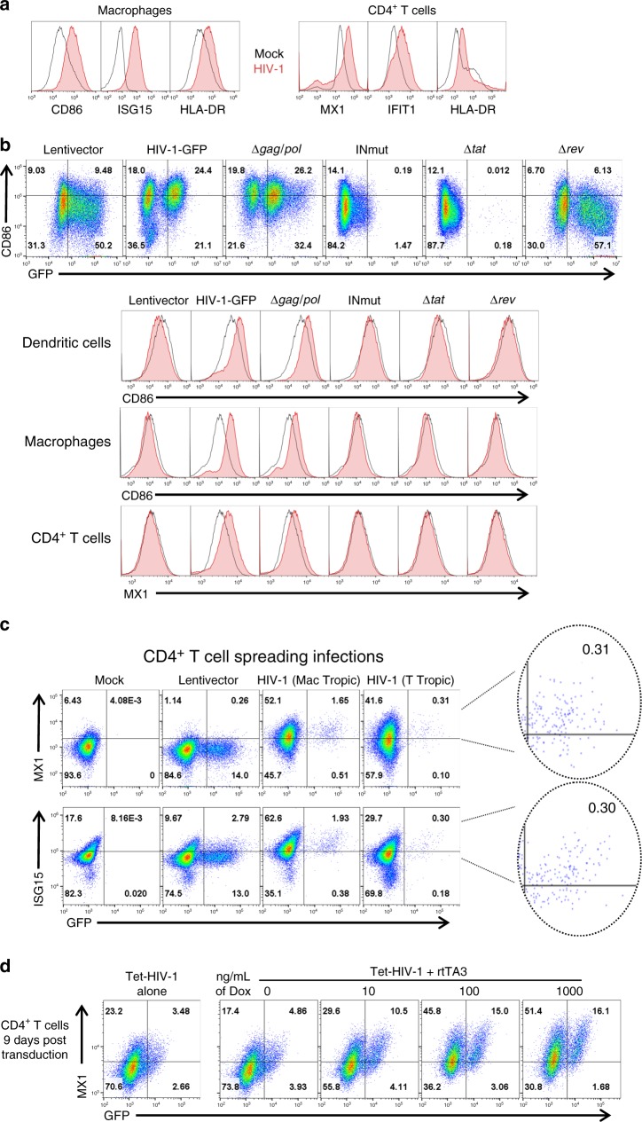
HIV-1-infected people who take drugs that suppress viremia to undetectable levels are protected from developing AIDS. Nonetheless, HIV-1 establishes proviruses in long-lived CD4+ memory T cells, and perhaps other cell types, that preclude elimination of the virus even after years of continuous antiviral therapy. Here we show that the HIV-1 provirus activates innate immune signaling in isolated dendritic cells, macrophages, and CD4+ T cells. Immune activation requires transcription from the HIV-1 provirus and expression of CRM1-dependent, Rev-dependent, RRE-containing, unspliced HIV-1 RNA. If rev is provided in trans, all HIV-1 coding sequences are dispensable for activation except those cis-acting sequences required for replication or splicing. Our results indicate that the complex, post-transcriptional regulation intrinsic to HIV-1 RNA is detected by the innate immune system as a danger signal, and that drugs which disrupt HIV-1 transcription or HIV-1 RNA metabolism would add qualitative benefit to current antiviral drug regimens.


Conflict of interest statement

The authors declare no competing interests.

Figures

Fig. 1
Fig. 1
HIV-1 transduction matures DCs. a Schematic of HIV-1-GFP, with frameshift in env (red line) and gfp in place of nef, and of the minimal lentivector, with self-inactivating ΔU3 LTR and gfp driven by the SFFV promoter. Unless indicated otherwise, vectors were pseudotyped with VSV G and cells were co-transduced with SIVMAC251 VLPs bearing Vpx. b Flow cytometry of DCs for GFP and CD86, after treatment as indicated. c Flow cytometry histograms for the indicated markers 72 h after DC transduction with HIV-1 (red) or mock (black). d Flow cytometry of DCs for GFP and CD86 after transduction with single-cycle clones, HIV-1NL4-3, HIV-1AD17, HIV-1Z331M-TF, or HIV-1ZM249M. e Transduction of DCs with HIV-2ROD-GFP, single-cycle vector. f Flow cytometry for CD86 and ISG15 of DCs treated for 24 h in the presence of nevirapine with a 1:1000 dilution of supernatant from autologous DCs transduced with the indicated vectors. g DC transduction with HIV-1-GFP in the absence of Vpx and the presence of 2 mM nucleosides. h 12-day spreading infection on DCs, with macrophage-tropic or T cell-tropic, replication-competent HIV-1, with or without SIV VLPs. i qRT-PCR quantitation of CXCL10 (black), IFNB1 (gray), or IL15 (white) mRNAs from DCs transduced with HIV-1-GFP. j qRT-PCR quantitation of CXCL10 mRNA in DCs transduced with either HIV-1-GFP or minimal lentivector, assessed at the indicated times post-transduction. k Cytokines in DC supernatant as assessed by luminex, 72 h after transduction with HIV-1-GFP (black) or minimal lentivector (gray). Shown are blood donor data representative of n = 12 (b), n = 4 (ck), or n = 8 (h). To determine significance, the MFI of all live cells for each sample was calculated as fold-change versus control. The exception being h where the MFI of only GFP+ cells was compared. When data from each donor replicate within a experiment was combined, the difference in MFI for all experimental vs control conditions was significant in all cases, p < 0.01; one-way ANOVA, Dunnett’s post-test. qRT-PCR and Luminex data were mean ± SD, p < 0.0001; two-way ANOVA, Dunnett’s post-test
Fig. 2
Fig. 2
Native HIV-1 RNA regulation is necessary for DC maturation. a Assessment of GFP and CD86 by flow cytometry following transduction with, top, HIV-1-GFP in the presence of 5 µM nevirapine (RTi), 10 µM raltegravir (INi), or no drug, and, bottom, HIV-1-GFP bearing mutant RT-D185K/D186L (RTmut) or mutant IN-D116A (INmut). b qRT-PCR quantitation of CXCL10 mRNA from the same DCs as a. c DCs treated with 1 µM of the TBK1 inhibitor BX795, or expressing shRNAs targeting either IRF3 or luciferase control, were challenged with 25 µg/mL cGAMP or HIV-1-GFP and assayed by flow cytometry for CD86 and ISG15. d Flow cytometry of DCs after transduction with minimal lentivectors expressing codon optimized HIV-1 genes; e, HIV-1-GFP in which translation was disrupted by two frameshifts in gag or by mutation of the first 14 AUGs in gag; f, HIV-1-GFP bearing deletion mutations encompassing gag/pol, vif/vpr, vpu/env, or nef/U3-LTR; g, HIV-1-GFP bearing mutations in tat or rev, co-transduced with both mutants, or co-transduced with minimal vector expressing tat and rev in trans; or h minimal lentivector with GFP in place of gag, SFFV promoter driving expression of tat, rev, or both, and repaired U3 in the 3′ LTR. When an essential viral component was disrupted within HIV-1-GFP, the factor in question was provided in trans, either during assembly in transfected HEK293 cells, or within transduced DCs, as appropriate (see Methods). Shown are blood donor data representative of n = 6 (a, b, e, f), n = 12 (c, g, h), n = 8 (d). To determine significance, the MFI of individual flow cytometry samples was calculated as fold-change versus control. When data from each donor replicate within a experiment were combined, the difference in MFI for all experimental vs control conditions was significant in all cases, p < 0.01; one-way ANOVA, Dunnett’s post-test against HIV-1-GFP for a, c, d, h or lentivector control for e, f. qRT-PCR data are mean±SD (p < 0.0001; two-way ANOVA, Dunnett’s post-test)
Fig. 3
Fig. 3
Rev-mediated RNA export is necessary for DC maturation but Tat is dispensable. a Optimized 2xTet operator was cloned into the 3’LTR of HIV-1-GFPΔtat to generate Tet-HIV-1; the strand-transfer reactions that occur during reverse transcription generate a Tet-regulated 5′-LTR in the provirus. DCs transduced with Tet-HIV-1, rtTA3, or both, were treated for 3 d with 500 ng/mL doxycycline and assayed by flow cytometry for p24, GFP, and CD86. b DCs co-transduced with Tet-HIV-1 and rtTA3 were treated with increasing concentrations of doxycycline. c To generate HIV-1-RTE/CTE, the RTEm26CTE element was cloned in place of nef in HIV-1-GFPΔrev/ΔRRE. DCs were transduced with the indicated vectors and assessed for p24 and ISG15 by flow cytometry. d DCs were treated with 25 nM leptomycin B, transduced with HIV-1-GFP, and assessed for GFP and ISG15 by flow cytometry. e DCs were treated with 25 nM leptomycin B, transduced with HIV-1-GFP or infected with Sendai virus (SeV), and assessed for ISG15 by flow cytometry. Shown are blood donor data representative of n = 10 (a, c), n = 4 (b), n = 6 (d, e). To determine significance, the MFI of individual flow cytometry samples was calculated as fold-change versus control. When data from each donor replicate within a experiment was combined, the difference in MFI for all experimental vs control conditions was significant in all cases, p < 0.01; one-way ANOVA, Dunnett’s post-test against dox negative control for a, b or HIV-1-GFP for ce
Fig. 4
Fig. 4
Innate immune activation in macrophages and CD4+ T cells. a Macrophages and CD4+ T cells were transduced with HIV-1-GFP and assayed 3 days later for the indicated activation markers. b DCs, macrophages, and CD4+ T cells were challenged with HIV-1-GFP or the indicated mutants. When an essential viral component was disrupted within HIV-1-GFP, the factor in question was provided in trans during assembly in transfected HEK293 cells, as appropriate (see Methods). The upper panel shows flow cytometry of the DCs for GFP and CD86. The histograms show CD86 for DCs and macrophages or MX1 for CD4+ T cells. c 12-day spreading infections on CD4+ T cells with either macrophage-tropic or T cell-tropic, replication-competent HIV-1. d CD4+ T cells were stimulated for 3 days with PHA and IL2, and transduced with Tet-HIV-1 and rtTA3. Cells were then cultured without stimulation for 9 days. Doxycycline was then added at the indicated concentrations. Cells were assayed for GFP and MX1 3 days later. Shown are blood donor data representative of n = 6 (a), n = 4 (bd). To determine significance, the MFI of individual flow cytometry samples was calculated as fold-change versus control. The exception being c where the MFI of only GFP+ cells was compared. When data from each donor replicate within a experiment were combined, the difference in MFI for all experimental vs control conditions was significant in all cases, p < 0.01; one-way ANOVA, Dunnett’s post-test against lentivector control for ac or dox control for d

Similar articles

Cited by

References

    1. Arts EJ, Hazuda DJ. HIV-1 antiretroviral drug therapy. Cold Spring Harb. Perspect. Med. 2012;2:a007161. doi: 10.1101/cshperspect.a007161. - DOI - PMC - PubMed
    1. Günthard HF, et al. Antiretroviral drugs for treatment and prevention of HIV infection in adults: 2016 recommendations of the International Antiviral Society-USA Panel. JAMA. 2016;316:191–210. doi: 10.1001/jama.2016.8900. - DOI - PMC - PubMed
    1. Kandathil AJ, Sugawara S, Balagopal A. Are T cells the only HIV-1 reservoir? Retrovirology. 2016;13:86. doi: 10.1186/s12977-016-0323-4. - DOI - PMC - PubMed
    1. Siliciano JD, et al. Long-term follow-up studies confirm the stability of the latent reservoir for HIV-1 in resting CD4+T cells. Nat. Med. 2003;9:727–728. doi: 10.1038/nm880. - DOI - PubMed
    1. Jiang AP, et al. Human mucosal mast cells capture HIV-1 and mediate viral trans-infection of CD4+T cells. J. Virol. 2015;90:2928–2937. doi: 10.1128/JVI.03008-15. - DOI - PMC - PubMed

Publication types

MeSH terms