Case Reports
. 2018 Jul-Dec;9(2):235-239.
doi: 10.4103/njms.NJMS_36_18.

Anchorage possibilities in case of a unilateral maxillary defect using the concept of Strategic Implant®

Affiliations
Case Reports

Anchorage possibilities in case of a unilateral maxillary defect using the concept of Strategic Implant®

Stefan Ihde et al. Natl J Maxillofac Surg. 2018 Jul-Dec.

Abstract

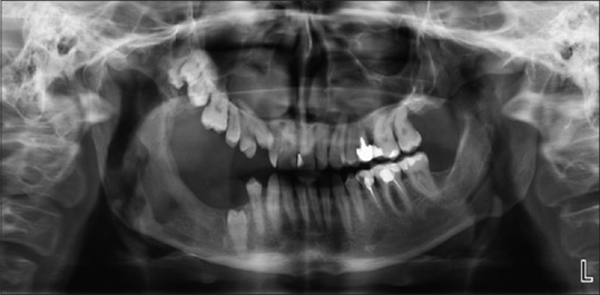
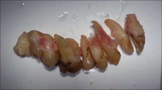
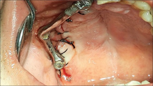
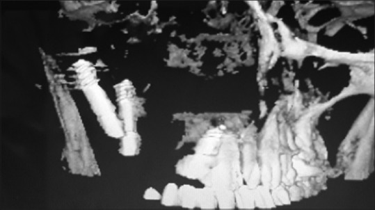
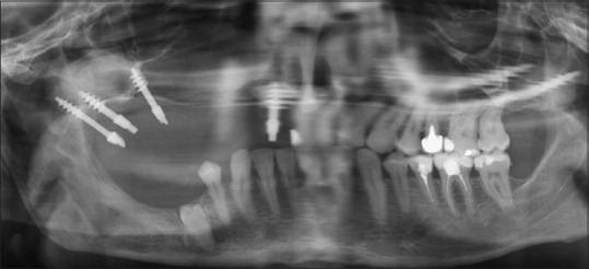
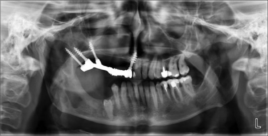
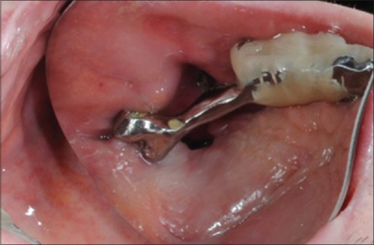
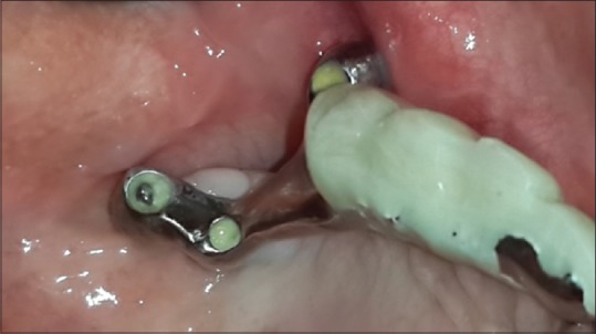
The aim of this article was to present the treatment method applied to a patient who suffered from a multiple fracture of the right maxilla, as a result of an accident that happened to him at an early age. The main consequence of this injury was an inhibited growth of the maxillary bone segment due to the lack of functional stimulus. The treatment consisted of four phases: the removal of all the teeth in the upper right maxilla, the immediate restoration with bicortical implants, the immediate prosthetic rehabilitation, and closing the oroantral communication.

Keywords: Cortical anchorage; Strategic Implant®; immediate loading; maxillary defect; supporting polygon.


Conflict of interest statement

There are no conflicts of interest.

Figures

Figure 1
Figure 1
Preoperative panoramic overview picture. Inhibited growth of the maxillary bone on the injured right side, missing teeth in the lower jaw with tooth 45 being partially retented. Due to the abnormal function, the morphology of the right temporomandibular joint had altered
Figure 2
Figure 2
(a) Clinical view on the dentition of the upper jaw before the intervention. The teeth are well in function from 11 to 27. All other upper teeth were in infraocclusion. The molars were hardly erupted and are therefore not visible here. (b) An intraoral clinical picture taken during the first treatment phase. During the extraction not only the teeth came out, but also the minimal rests of cortical bone surrounding them resulting in a large communication between the oral cavity and the maxillary sinus. The bone was partly missing up to the median raphe of the maxilla of the right side
Figure 3
Figure 3
All teeth in the upper-right jaw were extracted, from 11 to 19(!). It was decided that also the upper right central had to be removed, to make space for a stable, cortically anchored Strategic Implant®. This was necessary to set up a wide supporting polygon with masticatory surfaces in its center
Figure 4
Figure 4
A clinical picture after wound closure and immediate splinting of the three implants with a casted bar made from CoCrMo. This first construction (without the possibility of masticatory function) was left in place until we were able to place the 4th implant
Figure 5
Figure 5
The patient was sent to CT to determine the position of the 1st three implants placed and to search for more bone and determine the spatial relationship between the bone and the implants which were already placed
Figure 6
Figure 6
Panoramic overview picture after placement of the 4th implant (the 2nd implant in the tubero-pterygoid region)
Figure 7
Figure 7
Panoramic overview of the first prosthetic construction which allowed real masticatory function on the right side, but with reduced length of the chewing table
Figure 8
Figure 8
For several months, the patient had to accept a small but disturbing oroantral communication. After three unsuccessful attempts to close the tissue surgically, the condition suddenly healed without any surgery. We observed that the position of the communication gradually moved distally by itself before it finally closed
Figure 9
Figure 9
An overview on the first fixed, cemented reconstruction. All occlusal contacts are within the supporting polygon marked by two tubero-pterygoid screw implants, one implant in the caudal stump of the zygomatic bone, and one implant in the area of the right upper central with anchorage in the cortical floor of the nose.

References

    1. Wolff J. The Law of Bone Remodelling. In: Maquet P, Furlong R, translators. Verlag Von August Hirshwald. Verlag: Springer; 1892.
    1. Frost HM. Wolff's law and bone's structural adaptations to mechanical usage: An overview for clinicians. Angle Orthod. 1994;64:175–88. - PubMed
    1. Hanau RL. Articulation defined, analyzed and formulated. J Am Dent Assoc. 1926;13:1694–709.
    1. Planas P. Equilibrium and neuro-occlusal rehabilitation. Orthod Fr. 1992;63(Pt 2):435–41. - PubMed
    1. Dickerson WG, Chan CA, Carlson J. The human stomatognathic system: A scientific approach to occlusion. Dent Today. 2001;20:100. - PubMed

Publication types