. 2018 Nov 28:10:393.
doi: 10.3389/fnagi.2018.00393. eCollection 2018.

Predicting the Development of Normal-Appearing White Matter With Radiomics in the Aging Brain: A Longitudinal Clinical Study

Affiliations

Predicting the Development of Normal-Appearing White Matter With Radiomics in the Aging Brain: A Longitudinal Clinical Study

Yuan Shao et al. Front Aging Neurosci. .

Abstract

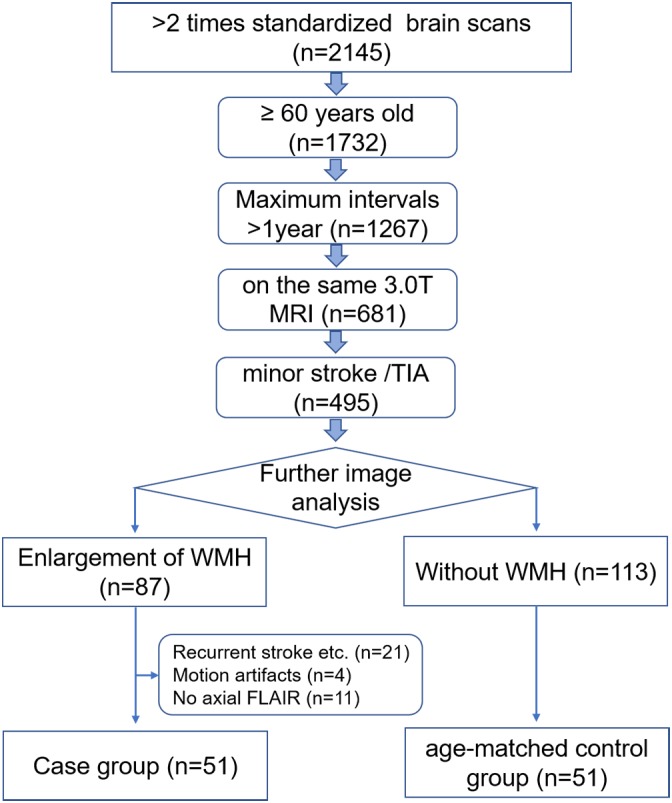
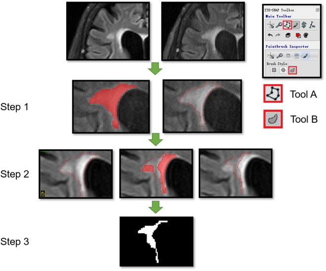
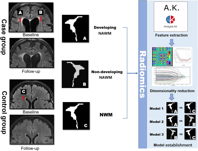
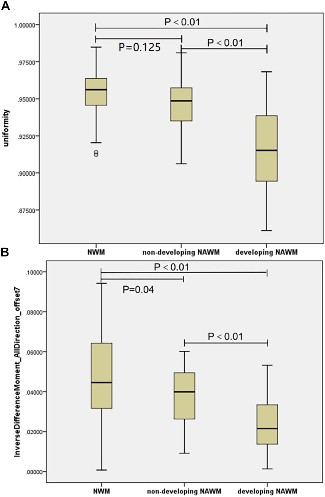
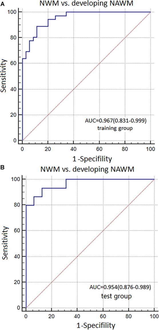
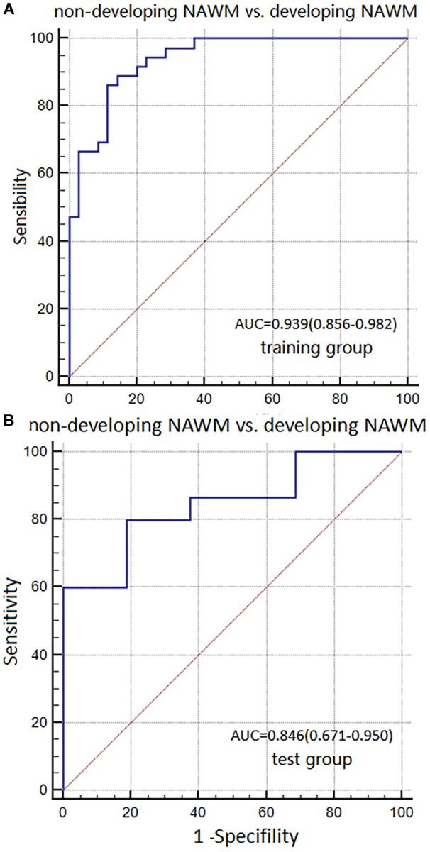
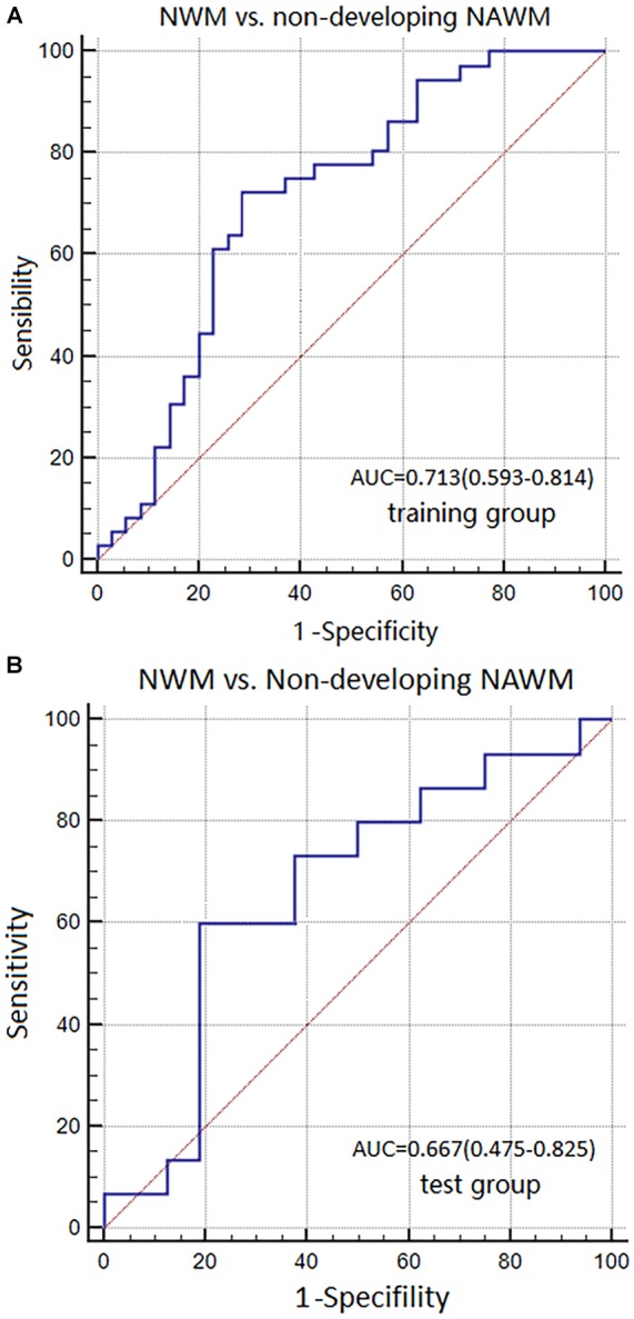
Background: Normal-appearing white matter (NAWM) refers to the normal, yet diseased tissue around the white matter hyperintensities (WMH) on conventional MR images. Radiomics is an emerging quantitative imaging technique that provides more details than a traditional visual analysis. This study aims to explore whether WMH could be predicted during the early stages of NAWM, using a textural analysis in the general elderly population. Methods: Imaging data were obtained from PACS between 2012 and 2017. The subjects (≥60 years) received two or more MRI exams on the same scanner with time intervals of more than 1 year. By comparing the baseline and follow-up images, patients with noted progression of WMH were included as the case group (n = 51), while age-matched subjects without WMH were included as the control group (n = 51). Segmentations of the regions of interest (ROIs) were done with the ITK software. Two ROIs of developing NAWM (dNAWM) and non-developing NAWM (non-dNAWM) were drawn separately on the FLAIR images of each patient. dNAWM appeared normal on the baseline images, yet evolved into WMH on the follow-up images. Non-dNAWM appeared normal on both the baseline and follow-up images. A third ROI of normal white matter (NWM) was extracted from the control group, which was normal on both baseline and follow-up images. Textural features were dimensionally reduced with ANOVA+MW, correlation analysis, and LASSO. Three models were built based on the optimal parameters of dimensional reduction, including Model 1 (NWM vs. dNAWM), Model 2 (non-dNAWM vs. dNAWM), and Model 3 (NWM vs. non-dNAWM). The ROC curve was adopted to evaluate the classification validity of these models. Results: Basic characteristics of the patients and controls showed no significant differences. The AUC of Model 1 in training and test groups were 0.967 (95% CI: 0.831-0.999) and 0.954 (95% CI: 0.876-0.989), respectively. The AUC of Model 2 were 0.939 (95% CI: 0.856-0.982) and 0.846 (95% CI: 0.671-0.950). The AUC of Model 3 were 0.713 (95% CI: 0.593-0.814) and 0.667 (95% CI: 0.475-0.825). Conclusion: Radiomics textural analysis can distinguish dNAWM from non-dNAWM on FLAIR images, which could be used for the early detection of NAWM lesions before they develop into visible WHM.

Keywords: FLAIR; MRI; longitudinal study; normal-appearing white matter (NAWM); radiomics; texture analysis; white matter hyperintensity.


Figures

FIGURE 1
FIGURE 1
Flowchart details the process of selecting the study subjects.
FIGURE 2
FIGURE 2
Segmentation of dNAWM in a 72-year old female patient. The time interval between the two images is 658 days. The WMH significantly progressed near the lateral ventricle forefoot. A and B tools from the ITK software were used. Tool A automatically covering the pixelated regions with similar gray levels and tool B being used to draw the outline of ROI and overlaying to other images. This process consisted of three steps: (1) using tools A and B to draw the outline of WMH on the follow-up images; (2) moving the ROI outline to the corresponding position on the baseline image and using tool A to cover the WMH on the baseline image; and (3) using tool B to modify the ROI boundaries and assessing the ROI segmentation by subtracting the baseline WMH from the follow-up WMH map.
FIGURE 3
FIGURE 3
Schematic diagram showing the research methods. dNAWM: appeared normal on FLAIR at the baseline, yet becomes WMH by the follow-up; non-dNAWM: appeared normal on both baseline and follow-up images; (normal white matter) NWM: considered as the standard of NWM. These three ROIs were segmented for feature extraction, dimensionality reduction, and model establishment.
FIGURE 4
FIGURE 4
The boxplots of co-occurring textural parameters among the NAWM, Non-dNAWM, and dNAWM in the (A) Uniformity and (B) IDM_AllDirection_offset7.
FIGURE 5
FIGURE 5
ROC curves were used to analyze the discriminatory power of Model 1 between the NWM and dNAWM in the (A) training group and (B) test group.
FIGURE 6
FIGURE 6
ROC curves were used to analyze the discriminatory power of Model 2 between the non-dNAWM and dNAWM in the (A) training group and (B) test group.
FIGURE 7
FIGURE 7
ROC curves were used to analyze the discriminatory power of Model 3 between the NWM and non-dNAWM in the (A) training group and (B) test group.

References

    1. Albuquerque M., Anjos L. G. V., Helen M. T. D. A., Oliveira M. S., Castellano G., Nucci A. (2015). MRI texture analysis reveals deep gray nuclei damage in amyotrophic lateral sclerosis. J. Neuroimaging 26 201–206. 10.1111/jon.12262 - DOI - PubMed
    1. Ardakani A. A., Gharbali A., Saniei Y., Mosarrezaii A., Nazarbaghi S. (2015). Application of texture analysis in diagnosis of multiple sclerosis by magnetic resonance imaging. Glob. J. Health Sci. 7 68–78. 10.5539/gjhs.v7n6p68 - DOI - PMC - PubMed
    1. Bae J. M., Ji Y. J., Lee H. Y., Sohn I., Kim H. S., Ji Y. S., et al. (2016). Pathologic stratification of operable lung adenocarcinoma using radiomics features extracted from dual energy CT images. Oncotarget 8 523–535. 10.18632/oncotarget.13476 - DOI - PMC - PubMed
    1. Barkovich A. J. (2000). Concepts of myelin and myelination in neuroradiology. Am. J. Neuroradiol. 21 1099–1109. - PMC - PubMed
    1. Bronge L., Bogdanovic N., Wahlund L. O. (2002). Postmortem MRI and histopathology of white matter changes in alzheimer brains. Dement. Geriatr. Cogn. Disord. 13 205–212. 10.1159/000057698 - DOI - PubMed

LinkOut - more resources