

Changes of Intracellular Porphyrin, Reactive Oxygen Species, and Fatty Acids Profiles During Inactivation of Methicillin-Resistant Staphylococcus aureus by Antimicrobial Blue Light
- PMID: 30546315
- PMCID: PMC6279940
- DOI: 10.3389/fphys.2018.01658
Changes of Intracellular Porphyrin, Reactive Oxygen Species, and Fatty Acids Profiles During Inactivation of Methicillin-Resistant Staphylococcus aureus by Antimicrobial Blue Light
Abstract
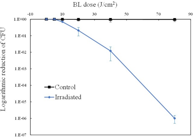
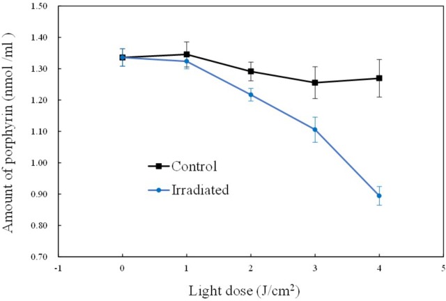
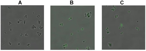
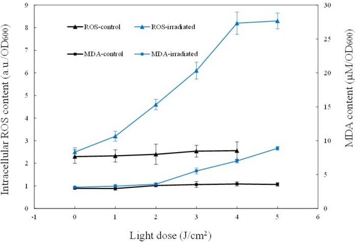
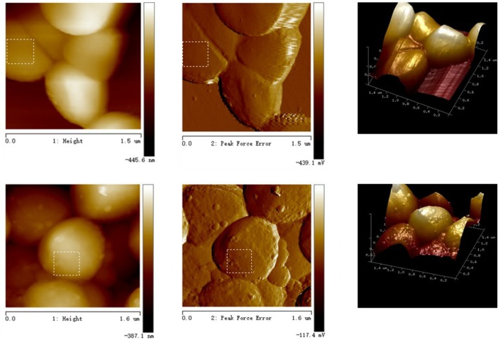
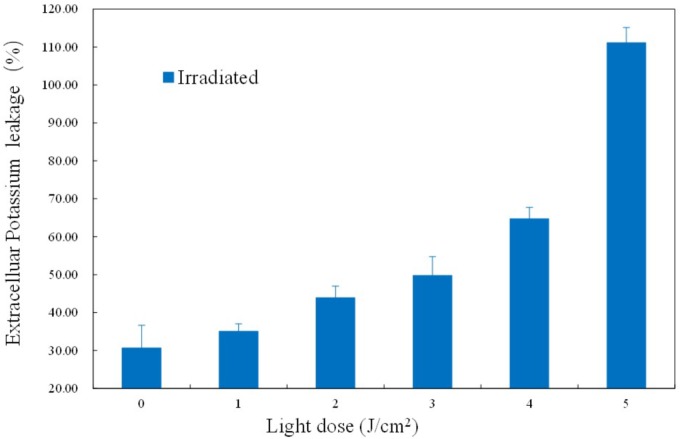
Antimicrobial blue light (aBL) has attracted increasing interest for its antimicrobial properties. However, the underlying bactericidal mechanism has not yet been verified. One hypothesis is that aBL causes the excitation of intracellular chromophores; leading to the generation of reactive oxygen species (ROS) and the resultant oxidization of various biomolecules. Thus, monitoring the levels of redox-sensitive intracellular biomolecules such as coproporphyrins, as well as singlet oxygen and various ROS may help to uncover the physiological changes induced by aBL and aid in establishing the underlying mechanism of action. Furthermore, the identification of novel targets of ROS, such as fatty acids, is of potential significance from a therapeutic perspective. In this study, we sought to investigate the molecular impact of aBL treatment on methicillin-resistant Staphylococcus aureus (MRSA). The results showed that aBL (5-80 J/cm2) exhibited a bactericidal effect on MRSA, and almost no bacteria survived when 80 J/cm2 had been delivered. Further studies revealed that the concentrations of certain intracellular molecules varied in response to aBL irradiation. Coproporphyrin levels were found to decrease gradually, while ROS levels increased rapidly. Moreover, imaging revealed the emergence and increase of singlet oxygen molecules. Concomitantly, the lipid peroxidation product malondialdehyde (MDA) increased in abundance and intracellular K+ leakage was observed, indicating permeability of the cell membrane. Atomic force microscopy showed that the cell surface exhibited a coarse appearance. Finally, fatty acid profiles at different illumination levels were monitored by GC-MS. The relative amounts of three unsaturated fatty acids (C16:1, C20:1, and C20:4) were decreased in response to aBL irradiation, which likely played a key role in the aforementioned membrane injuries. Collectively, these data suggest that the cell membrane is a major target of ROS during aBL irradiation, causing alterations to membrane lipid profiles, and in particular to the unsaturated fatty acid component.
Keywords: antimicrobial blue light; coproporphyrin; lipids; membrane injuries; methicillin-resistant Staphylococcus aureus; unsaturated fatty acids.
Figures
Similar articles
-
New Insights into the Bacterial Targets of Antimicrobial Blue Light.Microbiol Spectr. 2023 Feb 21;11(2):e0283322. doi: 10.1128/spectrum.02833-22. Online ahead of print. Microbiol Spectr. 2023. PMID: 36809152 Free PMC article.
-
Inactivation of Cronobacter sakazakii by blue light illumination and the resulting oxidative damage to fatty acids.Can J Microbiol. 2019 Dec;65(12):922-929. doi: 10.1139/cjm-2019-0054. Epub 2019 Sep 16. Can J Microbiol. 2019. PMID: 31525298
-
Berberine Damages the Cell Surface of Methicillin-Resistant Staphylococcus aureus.Front Microbiol. 2020 Apr 28;11:621. doi: 10.3389/fmicb.2020.00621. eCollection 2020. Front Microbiol. 2020. PMID: 32411101 Free PMC article.
-
Harnessing the power of light to treat staphylococcal infections focusing on MRSA.Curr Pharm Des. 2015;21(16):2109-21. doi: 10.2174/1381612821666150310102318. Curr Pharm Des. 2015. PMID: 25760339 Review.
-
Antimicrobial blue light: A 'Magic Bullet' for the 21st century and beyond?Adv Drug Deliv Rev. 2022 Jan;180:114057. doi: 10.1016/j.addr.2021.114057. Epub 2021 Nov 18. Adv Drug Deliv Rev. 2022. PMID: 34800566 Free PMC article. Review.
Cited by
-
Effectiveness of Ultra-High Irradiance Blue-Light-Emitting Diodes to Control Salmonella Contamination Adhered to Dry Stainless Steel Surfaces.Microorganisms. 2024 Jan 4;12(1):103. doi: 10.3390/microorganisms12010103. Microorganisms. 2024. PMID: 38257930 Free PMC article.
-
Low-Power NIR-Triggered Photothermal Inactivation of Pseudomonas aeruginosa with Polypyrrole Nanoparticles.Polymers (Basel). 2025 May 23;17(11):1442. doi: 10.3390/polym17111442. Polymers (Basel). 2025. PMID: 40508686 Free PMC article.
-
Antimicrobial Blue Light for Prevention and Treatment of Highly Invasive Vibrio vulnificus Burn Infection in Mice.Front Microbiol. 2022 Jul 12;13:932466. doi: 10.3389/fmicb.2022.932466. eCollection 2022. Front Microbiol. 2022. PMID: 35903474 Free PMC article.
-
Adjunctive effect of 470-nm and 630-nm light-emitting diode irradiation in experimental periodontitis treatment: a preclinical study.J Periodontal Implant Sci. 2024 Feb;54(1):13-24. doi: 10.5051/jpis.2203580179. Epub 2023 May 9. J Periodontal Implant Sci. 2024. PMID: 37336520 Free PMC article.
-
Mimicking the Effects of Antimicrobial Blue Light: Exploring Single Stressors and Their Impact on Microbial Growth.Antioxidants (Basel). 2024 Dec 23;13(12):1583. doi: 10.3390/antiox13121583. Antioxidants (Basel). 2024. PMID: 39765911 Free PMC article.
References
-
- Bumah V. V., Aboualizadeh E., Masson-Meyers D. S., Eells J. T., Enwemeka C. S., Hirschmugl C. J. (2017). Spectrally resolved infrared microscopy and chemometric tools to reveal the interaction between blue light (470nm) and methicillin-resistant Staphylococcus aureus. J. Photoch. Photobio. B 167 150–157. 10.1016/j.jphotobiol.2016.12.030 - DOI - PubMed
Grants and funding
LinkOut - more resources
Full Text Sources
Miscellaneous