

Persistent right superior vena cava in a patient with dextrocardia: Case report and review of the literature
- PMID: 30546510
- PMCID: PMC6281728
- DOI: 10.1016/j.jccase.2014.05.005
Persistent right superior vena cava in a patient with dextrocardia: Case report and review of the literature
Abstract
Introduction: Systemic venous circulation anomalies are uncommon; they are often incidental findings during echocardiography.
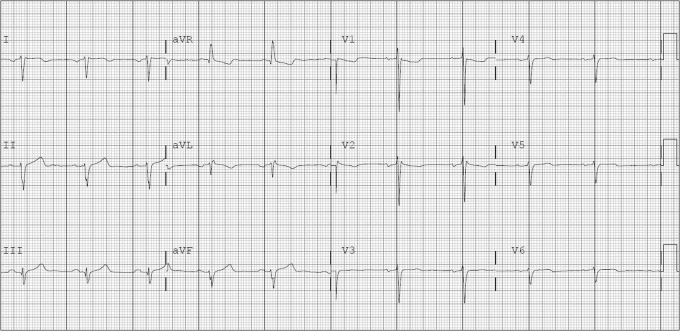
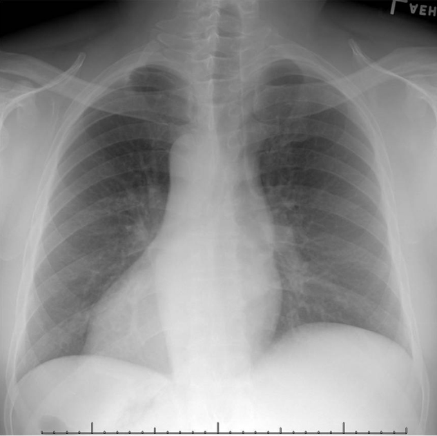
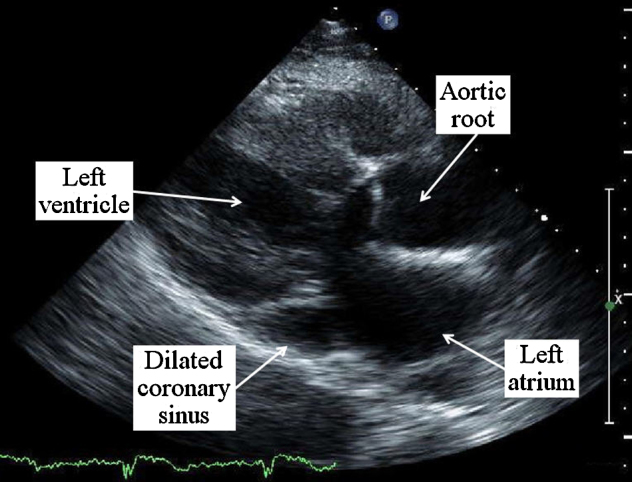
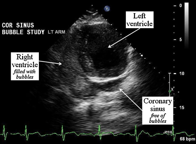
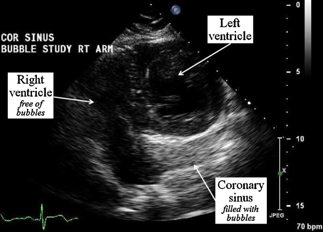
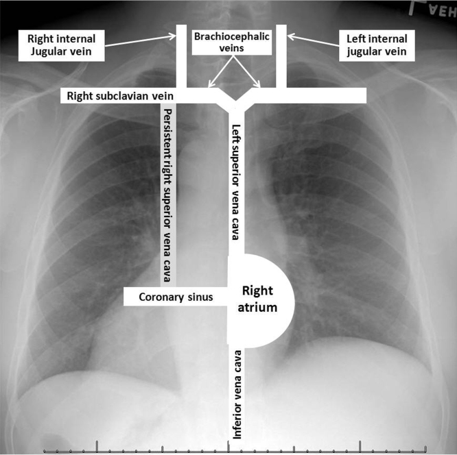
Case: A 56-year-old man, with dextrocardia, was evaluated for dyspnea. The patient's medical history included diabetes mellitus requiring insulin treatment, hypertension, and tobacco use. Physical examination revealed normal jugular venous pulsations and clear lungs. Cardiac examination revealed normal heart sounds, and grade II/VI systolic ejection murmur over the right precordium. Echocardiography revealed normal chamber size and systolic function, without significant valvular lesions. The coronary sinus was dilated. It was evaluated using intravenous agitated saline contrast to rule out anomalous venous drainage or shunting. When injected into the left antecubital vein, contrast appeared initially in the right atrium followed by the right ventricle. However, when injected into the right antecubital vein, contrast appeared initially in the dilated coronary sinus followed by the right atrium and right ventricle. There was no evidence of intracardiac shunting. These findings were consistent with persistent right superior vena cava in the setting of situs inversus dextrocardia, with normally draining left superior vena cava.
Conclusion: Persistent superior vena cava connection to the coronary sinus is often incidental but an important finding which helps in planning safe invasive procedures.<Learning objective: Understand the importance of identifying anomalous venous connections with regard to catheter-based procedures. Appreciate the incidence of these vascular anomalies in the normal population and in congenital heart disease. Understand how echocardiography with intravenous agitated saline contrast can be helpful in the diagnosis of such anomalous venous connections.>.
Keywords: Anomalous venous connections; Dextrocardia; Echocardiography.
Figures
References
-
- Petronzelli S., Patruno N., Pontillo D. Persistent left superior vena cava: diagnosis with saline contrast echocardiography. Heart. 2008;94:835. - PubMed
-
- Bhatti S., Hakeem A., Ahmad U., Malik M., Kosolcharoen P., Chang S. Persistent left superior vena cava (PLSVC) with anomalous left hepatic vein drainage into the right atrium: role of imaging and clinical relevance. Vasc Med. 2007;12:319–324. - PubMed
-
- Higgs A., Paris S., Potter F. Discovery of left-sided superior vena cava during central venous catheterization. Br J Anaesth. 1998;81:260–261. - PubMed
-
- Walpot J., Pasteuning W., Zwienen J. Persistent left superior vena cava diagnosed by bedside echocardiography. J Emerg Med. 2010;38:638–641. - PubMed
Publication types
LinkOut - more resources
Full Text Sources
