

A case of cardiac calcified amorphous tumor (cardiac CAT) causing acute embolism in right common iliac artery
- PMID: 30546536
- PMCID: PMC6279980
- DOI: 10.1016/j.jccase.2014.10.012
A case of cardiac calcified amorphous tumor (cardiac CAT) causing acute embolism in right common iliac artery
Abstract
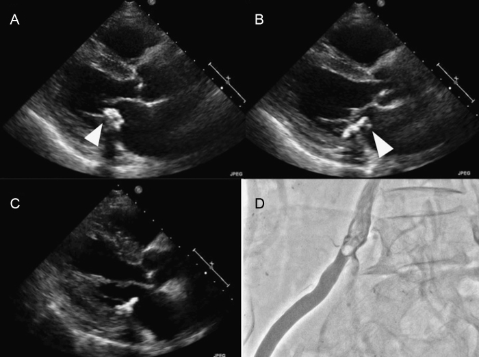
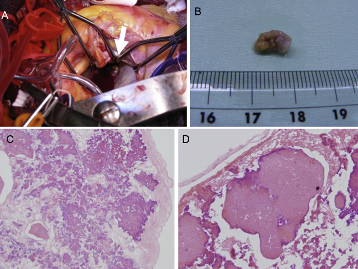
A 68-year-old man was admitted to our hospital for the further examination of intermittent claudication. He had been on continuous ambulatory peritoneal dialysis for 2 years. Screening transthoracic echocardiography (TTE) revealed a club-shaped tumor and a round-shaped tumor attached to mitral annulus calcification (MAC). The club-shaped tumor was swinging and plunged into the left ventricle at diastolic phase. Because of the risk of fatal embolism, we planned early surgical resection of the tumors. However, 13 days after admission, his intermittent claudication was getting worse and some part of the club-shaped tumor had vanished by TTE. Urgent iliac angiography showed that the tumor had embolized the right common iliac artery. Although we tried embolectomy using a Fogarty catheter, it was unsuccessful. We therefore treated the iliac artery stenosis by endovascular therapy and the procedure was successful. Three months later, he suffered from unstable angina and was treated by percutaneous coronary intervention. However, subacute stent thrombosis occurred after one month. After urgent treatment, we decided to treat him by coronary artery bypass graft and surgical resection of the residual tumor on MAC. The operation was performed successfully. Finally, the tumor was diagnosed as cardiac calcified amorphous tumor by its histologic features. <Learning objective: Cardiac calcified amorphous tumor (CAT) is a rare, non-neoplastic cardiac tumor. Mobile and pedunculated cardiac CAT is considered to be an important risk of systemic embolism. Based on our case and previous reports we reviewed cardiac CAT, especially MAC-related CAT, and it appears to be related to end-stage renal disease and may grow within a short duration. It is important to perform routine serial echocardiography for hemodialyzed patients in whom MAC has been identified.>.
Keywords: Acute embolism; Cardiac CAT; End-stage renal disease; MAC-related CAT; Thrombosis.
Figures
Similar articles
-
A rapidly growing cardiac calcified amorphous tumour diagnosed after coronary artery bypass graft surgery: a case report.Eur Heart J Case Rep. 2021 Aug 15;5(8):ytab243. doi: 10.1093/ehjcr/ytab243. eCollection 2021 Aug. Eur Heart J Case Rep. 2021. PMID: 34423238 Free PMC article.
-
Rapid growth of calcified amorphous tumor with mitral annulus calcification: a case report.Gen Thorac Cardiovasc Surg Cases. 2024 Aug 31;3(1):39. doi: 10.1186/s44215-024-00164-4. Gen Thorac Cardiovasc Surg Cases. 2024. PMID: 39517092 Free PMC article.
-
A case of cardiac calcified amorphous tumor complicated with acute myocardial infarction.J Cardiol Cases. 2022 Jan 31;25(6):396-399. doi: 10.1016/j.jccase.2022.01.004. eCollection 2022 Jun. J Cardiol Cases. 2022. PMID: 35685261 Free PMC article.
-
Intravascular Cooling Catheter-Related Venous Thromboembolism After Hypothermia: A Case Report and Review of the Literature.Ther Hypothermia Temp Manag. 2018 Jun;8(2):117-120. doi: 10.1089/ther.2017.0059. Epub 2018 Mar 23. Ther Hypothermia Temp Manag. 2018. PMID: 29570428 Review.
-
Particularities of a Cardiac Amorphous Left Ventricular Tumor in a Patient with Coronary Artery Disease-Diagnostic and Therapeutic Challenges: A Case Report and Literature Review.J Clin Med. 2024 Oct 12;13(20):6092. doi: 10.3390/jcm13206092. J Clin Med. 2024. PMID: 39458043 Free PMC article. Review.
Cited by
-
Calcified amorphous tumor located on a severely calcified mitral annulus in a patient with normal renal function.J Surg Case Rep. 2022 Jan 21;2022(1):rjab608. doi: 10.1093/jscr/rjab608. eCollection 2022 Jan. J Surg Case Rep. 2022. PMID: 35079337 Free PMC article.
-
A rapidly growing cardiac calcified amorphous tumour diagnosed after coronary artery bypass graft surgery: a case report.Eur Heart J Case Rep. 2021 Aug 15;5(8):ytab243. doi: 10.1093/ehjcr/ytab243. eCollection 2021 Aug. Eur Heart J Case Rep. 2021. PMID: 34423238 Free PMC article.
-
Cardiac calcified amorphous tumour associated with multiple myeloma.BMJ Case Rep. 2020 Apr 28;13(4):e233679. doi: 10.1136/bcr-2019-233679. BMJ Case Rep. 2020. PMID: 32350053 Free PMC article.
References
-
- Reynolds C., Tazelaar H.D., Edwards W.D. Calcified amorphous tumor of the heart (cardiac CAT) Hum Pathol. 1997;28:601–606. - PubMed
-
- Morishima A., Sasahashi N., Ueyama K. Calcified amorphous tumors with excision in hemodialysis patient: report of 2 cases. Kyobu Geka. 2006;59:851–854. - PubMed
-
- Kubota H., Fujioka Y., Yoshino H., Koji H., Yoshihara K., Tonari K., Endo H., Tsuchiya H., Mera H., Soga Y., Taniai S., Sakata K., Sudo K. Cardiac swinging calcified amorphous tumors in end-stage renal failure patients. Ann Throrac Surg. 2010;90:1692–1694. - PubMed
-
- Fujiwara M., Watanabe H., Iino T., Kobukai Y., Ishibashi K., Yamamoto H., Iino K., Yamamoto F., Ito H. Two cases of cardiac calcified amorphous tumor mimicking mitral valve vegetation. Circulation. 2012;125:e432–e434. - PubMed
-
- Nishigawa K., Takiuchi H., Kubo Y., Masaki H., Yanemoto K. Calcified amorphous tumor: three dimensional transesophageal echocardiography. Asian Cardiovasc Thorac Ann. 2012;20:355. - PubMed
Publication types
LinkOut - more resources
Full Text Sources
Other Literature Sources
Miscellaneous
