

Serial endovascular assessment of polytetrafluoroethylene-covered stent: Capabilities and limitations of intravascular imaging modalities affected by a temporal factor
- PMID: 30546539
- PMCID: PMC6279971
- DOI: 10.1016/j.jccase.2014.11.004
Serial endovascular assessment of polytetrafluoroethylene-covered stent: Capabilities and limitations of intravascular imaging modalities affected by a temporal factor
Abstract
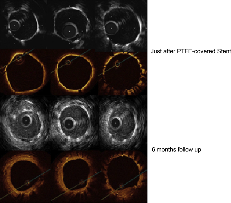
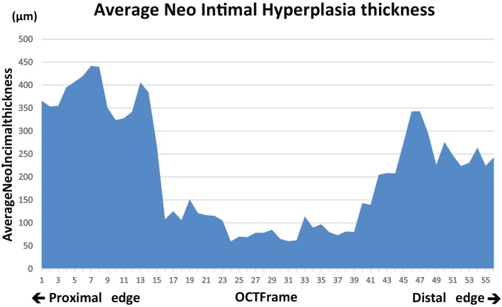
A 47-year-old male who previously underwent coronary bypass graft surgery was transferred to our hospital for treatment of bare metal in-stent restenosis (ISR) of severely calcified left main (LM) coronary lesion. During a repeat coronary intervention, LM coronary perforation occurred after rotational atherectomy followed by balloon dilatation. Hemostasis was successfully achieved by implantation of a single polytetrafluoroethylene (PTFE)-covered stent. Although intravascular ultrasound (IVUS) and optical coherence tomography (OCT) were documented, any additional information was not obtained except stent expansion. Routine 6-month follow-up angiography revealed no findings of restenosis. Three representative imaging modalities, IVUS, OCT, and angioscopy were applied to visualize and differentiate any structures within the PTFE-covered stent. Intravascular findings included, (1) vascular structures outside the covered stent could be observed sufficiently by both IVUS and OCT at this time that could not be seen at all just after implantation, (2) neointimal hyperplasia distributed dominantly at both stent edges, and (3) in-stent micro thrombi still existed even 6 months after implantation. Intravascular findings of PTFE-covered stent may vary between the observational periods. Furthermore, vascular healing process of this special stent may be different from those of non-covered mesh stents. <Learning objective: Even with the use of IVUS and OCT, it may be difficult to evaluate apposition of PTFE-covered stent just after implantation. However, it could be visualized as being sufficiently similar to the other common stents at 6-month follow-up. Unique longitudinal NIH distribution (bilateral edge dominant) was evaluated, and existence of micro thrombi within PTFE-covered stent even at 6 months.>.
Keywords: Angioscopy; IVUS; OCT; Polytetrafluoroethylene-covered stent.
Figures


Similar articles
-
Restenosis of a Polytetrafluoroethylene-Covered Stent Visualized by Coronary Angioscopy and Optical Coherence Tomography: A Case Report.Int J Angiol. 2020 Mar;29(1):58-62. doi: 10.1055/s-0039-1685510. Epub 2019 Apr 16. Int J Angiol. 2020. PMID: 32132819 Free PMC article.
-
Clinical and procedure characteristics in patients treated with polytetrafluoroethylene-covered stents after coronary perforation: a CIRC-8U multicenter registry and literature review.Cardiovasc Interv Ther. 2021 Oct;36(4):418-428. doi: 10.1007/s12928-020-00716-9. Epub 2020 Oct 9. Cardiovasc Interv Ther. 2021. PMID: 33037569 Review.
-
Six months clinical, angiographic, and IVUS follow-up after PTFE graft stent implantation in native coronary arteries.Acta Cardiol. 2000 Aug;55(4):255-60. doi: 10.2143/AC.55.4.2005748. Acta Cardiol. 2000. PMID: 11041124 Clinical Trial.
-
Covered stent implantation for calcified nodule to physically hinder its protrusion causing restenosis: a case report.Cardiovasc Diagn Ther. 2024 Oct 31;14(5):974-981. doi: 10.21037/cdt-24-216. Epub 2024 Oct 22. Cardiovasc Diagn Ther. 2024. PMID: 39513136 Free PMC article.
-
[New methods of coronary imaging II. Intracoronary ultrasonography in clinical practice].Ital Heart J Suppl. 2001 Jun;2(6):579-92. Ital Heart J Suppl. 2001. PMID: 11460831 Review. Italian.
Cited by
-
Clinical Outcomes following Large Vessel Coronary Artery Perforation Treated with Covered Stent Implantation: Comparison between Polytetrafluoroethylene- and Polyurethane-Covered Stents (CRACK-II Registry).J Clin Med. 2021 Nov 21;10(22):5441. doi: 10.3390/jcm10225441. J Clin Med. 2021. PMID: 34830722 Free PMC article.
-
A case of a coronary covered stent for repeated restenosis at the anastomosis site between saphenous vein graft and graft prosthesis.J Cardiol Cases. 2021 Aug 16;25(2):110-114. doi: 10.1016/j.jccase.2021.07.007. eCollection 2022 Feb. J Cardiol Cases. 2021. PMID: 35079311 Free PMC article.
-
Efficacy and Safety of a Polytetrafluoroethylene Membrane Wrapped a Single Layer of Sirolimus-Eluting Stent in a Porcine Coronary Perforation Model.Rev Cardiovasc Med. 2022 Jun 24;23(7):233. doi: 10.31083/j.rcm2307233. eCollection 2022 Jul. Rev Cardiovasc Med. 2022. PMID: 39076929 Free PMC article.
-
Mid-Term Outcomes of Novel Covered Stent with Biodegradable Membrane in Porcine Coronary Artery Perforation.Rev Cardiovasc Med. 2023 Jul 12;24(7):197. doi: 10.31083/j.rcm2407197. eCollection 2023 Jul. Rev Cardiovasc Med. 2023. PMID: 39077012 Free PMC article.
References
-
- Takano M., Yamamoto M., Murakami D., Inami S., Okamatsu K., Seimiya K., Ohba T., Seino Y., Mizuno K. Lack of association between large angiographic late loss and low risk of in-stent thrombus: angioscopic comparison between paclitaxel- and sirolimus-eluting stents. Circ Cardiovasc Interv. 2008;1:20–27. - PubMed
-
- Maehara A., Minz G.S., Lansky A.J., Witzenbickler B., Guagliumi G., Brodie B., Kellet M.A., Parise H., Mehran R., Stone G.W. Volumetric intravascular ultrasound analysis of paclitaxel-eluting and bare metal stents in acute myocardial infarction: the harmonizing outcomes with revascularization and stents in acute myocardial infarction intravascular ultrasound substudy. Circulation. 2009;120:1875–1882. - PubMed
-
- Gonzalo N., Barlis P., Serruys P.W., Garcia-Garcia H.M., Onuma Y., Ligthart J., Regar E. Incomplete stent apposition and delayed tissue coverage are more frequent in drug-eluting stents implanted during primary percutaneous coronary intervention for ST-segment elevation myocardial infarction than in drug-eluting stents implanted for stable/unstable angina: insights from optical coherence tomography. JACC Cardiovasc Interv. 2009;2:445–452. - PubMed
-
- Tanigawa J., Barlis P., Di Mario C. Intravascular optical coherence tomography: optimisation of image acquisition and quantitative assessment of stent strut apposition. EuroIntervention. 2007;3:128–136. - PubMed
-
- Dutary J., Zakhem B., DE Lucas C.B., Paulo M., Gonzalo N., Alfonso F. Treatment of a giant coronary artery aneurysm: intravascular ultrasound and optical coherence tomography findings. J Interv Cardiol. 2012;25:82–85. - PubMed
Publication types
LinkOut - more resources
Full Text Sources
Other Literature Sources
