

Primary cardiac malignant fibrous histiocytoma with abdominal wall metastasis
- PMID: 30546578
- PMCID: PMC6281857
- DOI: 10.1016/j.jccase.2015.05.014
Primary cardiac malignant fibrous histiocytoma with abdominal wall metastasis
Abstract
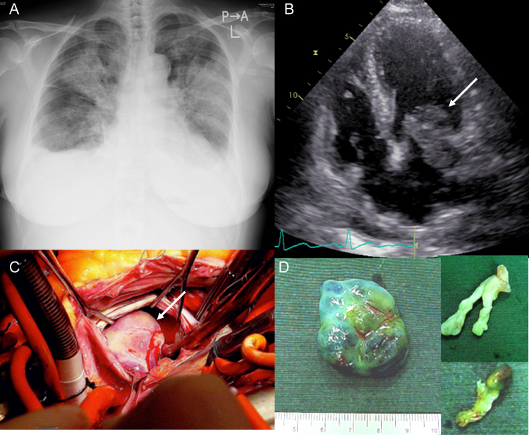
We present a rare case of cardiac malignant fibrous histiocytoma (MFH; undifferentiated pleomorphic sarcoma); to date, fewer than 100 cases of cardiac MFH have been reported. In this case, transthoracic echocardiography revealed cardiac tumors in the left atrium (LA) of a 53-year-old woman with a 3-month history of worsening dyspnea; the largest tumor was found to protrude through the mitral valve in diastole, causing stenosis. Three of the four tumors were resected during emergency surgery; however, the residual tumor extension into the left pulmonary vein could not be removed. Histological findings of the resected tumors, such as organized thrombus and myxomatous tissue changes, indicated that the tumors were benign. After 3 months, the patient underwent total resection for a small mass that developed on her right abdominal wall, which was revealed histologically to be MFH; additionally, the residual mass in the LA had enlarged progressively. After undergoing radiation therapy without further surgery, she died of cerebral bleeding 6 months after cardiac surgery. Postmortem examination revealed that the tumor in the LA was an MFH. Thus, cardiac MFH should be considered as a differential diagnosis for tumors on the posterior wall of the LA. <Learning objective: Primary cardiac malignant fibrous histiocytoma (MFH), which is easily mistaken for atrial myxoma, is a rare type of cardiac sarcoma. MFH occurs most commonly on the posterior wall of the left atrium (LA), and total resection is currently the only effective therapy; however, the prognosis is poor. Therefore, a high level of suspicion is required to facilitate early diagnosis. Cardiac MFH should be considered as a differential diagnosis for tumors on the posterior wall of the LA.>.
Keywords: Cardiac tumor; Malignant fibrous histiocytoma; Undifferentiated pleomorphic sarcoma.
Figures


References
-
- O’Brien J.E., Stout A.P. Malignant fibrous xanthomas. Cancer. 1964;17:1445–1455. - PubMed
-
- Okamoto K., Kato S., Katsuki S., Wada Y., Toyozumi Y., Morimatsu M., Aoyagi S., Imaizumi T. Malignant fibrous histiocytoma of the heart: case report and review of 46 cases in the literature. Intern Med. 2001;40:1222–1226. - PubMed
-
- Reynen K. Frequency of primary tumors of the heart. Am J Cardiol. 1996;77:107. - PubMed
-
- Simpson L., Kumar S.K., Okuno S.H., Schaff H.V., Porrata L.F., Buckner J.C., Moynihan T.J. Malignant primary cardiac tumors: review of a single institution experience. Cancer. 2008;112:2440–2446. - PubMed
-
- Shah A.A., Churg A., Sbarbaro J.A., Sheppard J.M., Lamberti J. Malignant fibrous histiocytoma of the heart presenting as an atrial myxoma. Cancer. 1978;42:2466–2471. - PubMed
LinkOut - more resources
Full Text Sources
Other Literature Sources
