. 2015 Nov 14;13(1):25-30.
doi: 10.1016/j.jccase.2015.09.004. eCollection 2016 Jan.

Starfix lead extraction: Clinical experience and technical issues

Affiliations

Starfix lead extraction: Clinical experience and technical issues

Pier Giorgio Golzio et al. J Cardiol Cases. .

Abstract

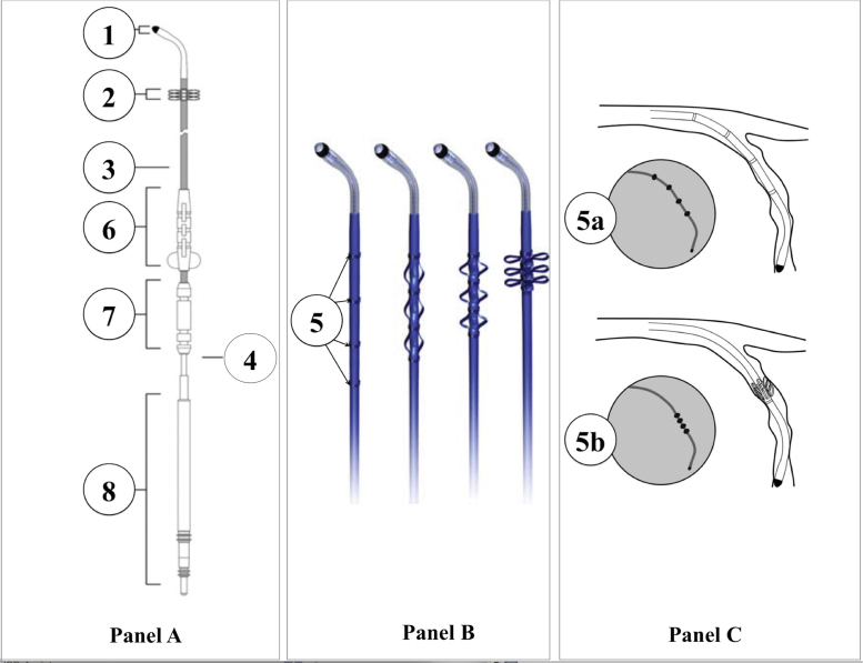
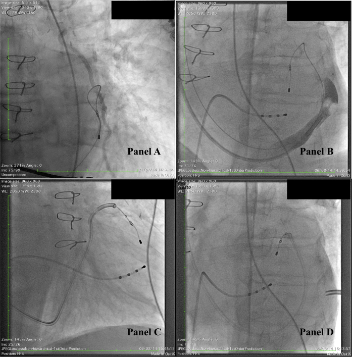
Transvenous lead extraction (TLE) of the Starfix coronary sinus (CS) active-fixation lead may be challenging, due to undeployment of fixation lobes and venous occlusion. We report our experience in Starfix TLE, in comparison with previous data. A 78-year-old male, implanted in 2009 with Starfix lead, was referred to our institution for TLE, due to infective endocarditis with lead-associated vegetations. The tip of Starfix lead was located in distant, anterior position, in the great cardiac vein, close to patent left internal mammary artery-to-left anterior descending artery anastomosis, and first-choice surgical removal had a prohibitive operative risk. Conventional dilatation beyond CS ostium, as well as the use of a standard delivery catheter, was ineffective. An off-label modification of the delivery, by cutting the distal soft tip, was successful. However, the tip of the lead fragmented and was trapped in the innominate vein. Then a gooseneck snare grasped the fragment, allowing complete retrieval. TLE of Starfix leads may be particularly challenging, especially when its tip is located in a distant anterior location. In these cases, important help may be obtained by dilatation within the CS, by means of conventional or modified delivery catheters. Only experienced operators, sometimes with non-conventional techniques, should perform TLE of Starfix leads. <Learning objective: TLE of Starfix leads may be challenging, particularly when the tip is located in a distant anterior position. Dilatation with conventional tools may be precluded. In these cases modifications of the delivery catheters may be useful. Surgery should be avoided as first-choice procedure; only experienced operators, sometimes with non-conventional techniques, should perform TLE of Starfix leads.>.

Keywords: Active-fixation leads; Coronary sinus; Infection; Lead extraction; Starfix lead.


Figures

Fig. 1
Fig. 1
Technical representation of the Starfix lead, with fixation lobes undeployed and deployed. Considering the anatomy of the coronary sinus, understanding how the fixation mechanism acts, and how deployed fixation lobes can cause occlusion of the coronary sinus, is easily allowed. Fibrosis and adherences can further ensue, so making lead extraction very risky and difficult. You can see the steroid-eluting tip (Panel A, 1), followed by three series of four polyurethane lobes each (A, 2). They can be deployed by advancing the push tubing along the lead (A, 3), so increasing the external diameter from the 5-French caliber of the lead body (A, 4) up to up to 24-French. Four radiopaque platinum–iridium indicator rings (Panel B, 5) on each side of the series of lobes can be seen under fluoroscopy to mark the extent of lobe undeployment (Panel C, 5a) and deployment (Panel C, 5b). Also shown in scheme, Panel A: standard fixation sleeve (6), lead sewing sleeve (7) and IS-1 unipolar connector (8). Panel B shows schematic representation of progressive deployment of fixation lobes, and Panel C the scheme of the Starfix lead within the target venous branch of the coronary sinus, immediately after attainment of the target vein, with lobes undeployed (5a, fluoro scheme within the circle) and deployed (5b, fluoro scheme within the circle). The pullback of the tubing should be able to undeploy lobes, but frequent drawbacks occur, due to failure of the mechanism itself, and/or fibrosis within and around lobes.
Fig. 2
Fig. 2
Panel A: coronary angiography. Selective angiography of left internal mammary artery (LIMA). LIMA-to-left anterior descending artery anastomosis is patent, and well working. This anastomosis ensures the perfusion to the viable myocardium, since proximal interventricular, circumflex, and right coronary arteries are occluded. This patient suffered an inferior myocardial infarction in 1991; myocardial perfusion with 99mTc-methoxyisobutylisonitrile stress/rest single-photon emission computed tomography disclosed a wide irreversible inferior and infero-lateral defect, and a small partially reversible apical defect. Panel B: selective retrograde coronary sinus (CS) venography, with a balloon-occluding Swan-Ganz catheter. The CS is cannulated through a conventional straight 50-cm long 7-French CS delivery (Attain Command CS Cannulation Catheter, Model 6250V-50S, Medtronic). After inflation of the Swan-Ganz balloon, occlusion of the mid coronary sinus is observed. Panel C: a 57-cm long 7-French straight CS delivery (Attain Command CS Cannulation Catheter, Model 6250VI-57S, Medtronic) is advanced over the left ventricular lead. The distal portion of the CS is reached, just before the angulation of the CS into the interventricular grove. This obtains a partial undeployment of the proximal series of fixation lobes. Panel D: off-label modification of the delivery, by cutting the soft distal collar of the catheter. The modification produces a greater pushing force along the lead, so reaching the origin of the great cardiac vein. The distal end of this modified delivery is firmly anchored to the proximal lobes.
Fig. 3
Fig. 3
Panel A: during extraction, the tip of the Starfix, not protected inside a dilator, is trapped into the innominate vein, just before the costo-clavicular angle narrowing. Manual traction results in fragmentation of the lead, with its tip retained. Due to the traction forces carried out, the fixation lobes are forced against the vessel wall, and again fully deployed, so causing venous occlusion. Panels B and C: an Amplatz Goose Neck 6-French snare catheter, with a 30 mm loop snare (ev3 Inc., Plymouth, MN, USA) catches the tip of the Starfix lead. Panel D: the traction over the tip undeploys the lobes, as can be seen at the mid innominate vein level. Panel E: the tip fragment of the Starfix lead is extracted through a long 18-French femoral sheath. Panel F: the tip Starfix fragment extracted.

References

    1. Nagele H., Azizi M., Hashagen S., Castel M.A., Behrens S. First experience with a new active fixation coronary sinus lead. Europace. 2007;9:437–441. - PubMed
    1. Crossley G.H., Exner D., Mead R.H., Sorrentino R.A., Hokanson R., Li S., Adler S. Chronic performance of an active fixation coronary sinus lead. Heart Rhythm. 2010;7:472–478. - PubMed
    1. Wilkoff B.L., Love C.J., Byrd C.L., Bongiorni M.G., Carrillo R.G., Crossley G.H., 3rd, Epstein L.M., Friedman R.A., Kennergren C.E., Mitkowski P., Schaerf R.H., Wazni O.M. Transvenous Lead extraction: Heart Rhythm Society expert consensus on facilities, training, indications, and patient management: this document was endorsed by the American Heart Association (AHA) Heart Rhythm. 2009;6:1085–1104. - PubMed
    1. Golzio P.G., Gabbarini F., Anselmino M., Vinci M., Gaita F., Bongiorni M.G. Gram-positive occult bacteremia in patients with pacemaker and mechanical valve prosthesis: a difficult therapeutic challenge. Europace. 2010;12:999–1002. - PubMed
    1. Golzio P.G., Manganiello S., Gaita F. Labelled leucocyte scintigraphy in an infected externalized Riata lead. Europace. 2014;16:1442. - PubMed