

Nonobstructive angioscopy in patient with atherosclerotic renal artery stenosis
- PMID: 30546775
- PMCID: PMC6281555
- DOI: 10.1016/j.jccase.2013.08.014
Nonobstructive angioscopy in patient with atherosclerotic renal artery stenosis
Abstract
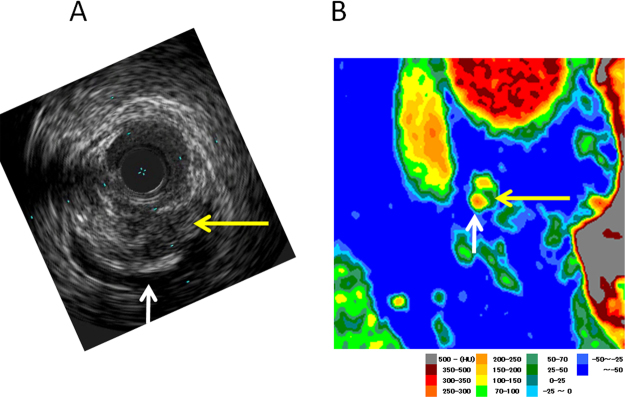
Few applications of angioscopy for evaluating atherosclerosis of the abdominal aorta have been described. We report the demonstration of atherosclerotic yellow plaque by nonobstructive angioscopy in a patient with left renal artery stenosis. Computed tomography angiography showed stenosis in one of the left renal arteries in a 65-year-old man who presented with renal impairment and hypertension. Invasive selective renal angiography indicated severe stenosis in the proximal portion of the inferior left renal arteries. Intravascular ultrasound demonstrated eccentric plaque with predominant low-density plaque with calcification as the culprit. Percutaneous transluminal renal angioplasty with stent implantation of the left renal artery was performed. Nonobstructive angioscopy demonstrated a grade 3 yellow culprit plaque at the proximal end of the stent, and grade 2 and grade 1 yellow plaques as the culprit plaques at the middle and distal portions of the artery, respectively. <Learning objective: Atherosclerotic renal artery stenosis characterized by lipid-rich plaque and yellow plaque was diagnosed by intravascular imaging, such as intravascular ultrasound and angioscopy. As the stenosis was hemodynamically significant, percutaneous transluminal renal angioplasty was successfully performed. Nonobstructive angioscopy may be potentially applied for monitoring of transluminal ablation of the renal artery sympathetic nerves during drug-resistant hypertension.>.
Keywords: Computed tomography angiography; Intravascular ultrasound; Nonobstructive angioscopy; Renal artery stenosis; Yellow plaque.
Figures


Similar articles
-
Correlation between plaque vulnerability of aorta and coronary artery: an evaluation of plaque activity by direct visualization with angioscopy.Int J Cardiovasc Imaging. 2015 Aug;31(6):1107-14. doi: 10.1007/s10554-015-0669-z. Epub 2015 Apr 28. Int J Cardiovasc Imaging. 2015. PMID: 25916323 Free PMC article.
-
In-Stent Yellow Plaque at 1 Year After Implantation Is Associated With Future Event of Very Late Stent Failure: The DESNOTE Study (Detect the Event of Very late Stent Failure From the Drug-Eluting Stent Not Well Covered by Neointima Determined by Angioscopy).JACC Cardiovasc Interv. 2015 May;8(6):814-821. doi: 10.1016/j.jcin.2014.12.239. JACC Cardiovasc Interv. 2015. PMID: 25999104
-
Plaque characterization and atherosclerosis evaluation by coronary angioscopy.Herz. 2003 Sep;28(6):501-4. doi: 10.1007/s00059-003-2486-8. Herz. 2003. PMID: 14569391
-
[New imaging methods for visualizing coronary arteries].Z Kardiol. 1998;87 Suppl 2:61-73. doi: 10.1007/s003920050540. Z Kardiol. 1998. PMID: 9827463 Review. German.
-
[The role of angioscopy and intravascular ultrasound imaging in acute coronary syndrome].J Cardiol. 1999 Mar;33 Suppl 1:17-21. J Cardiol. 1999. PMID: 10342132 Review. Japanese.
Cited by
-
Seeing is believing - Imaging of a plaque in the renal artery.J Cardiol Cases. 2013 Dec 13;9(2):84-85. doi: 10.1016/j.jccase.2013.09.011. eCollection 2014 Feb. J Cardiol Cases. 2013. PMID: 30534303 Free PMC article. No abstract available.
-
Comparison of angioscopy and histopathology for the evaluation of carotid plaque characteristics: an ex vivo validation study.Int J Cardiovasc Imaging. 2020 Feb;36(2):231-239. doi: 10.1007/s10554-019-01720-8. Epub 2019 Oct 29. Int J Cardiovasc Imaging. 2020. PMID: 31664681
References
-
- White C.J., Olin J.W. Diagnosis and management of atherosclerotic renal artery stenosis: improving patient selection and outcomes. Nat Clin Pract Cardiovasc Med. 2009;6:176–190. - PubMed
-
- Dubel G.J., Murphy T.P. The role of percutaneous revascularization for renal artery stenosis. Vasc Med. 2008;13:141–156. - PubMed
-
- Komatsu S., Hirayama A., Omori Y., Ueda Y., Mizote I., Fujisawa Y., Kiyomoto M., Higashide T., Kodama K. Detection of coronary plaque by computed tomography with a novel plaque analysis system, ‘Plaque Map’, and comparison with intravascular ultrasound and angioscopy. Circ J. 2005;69:72–77. - PubMed
-
- Hirayama A., Saito S., Ueda Y., Takayama T., Honye J., Komatsu S., Yamaguchi O., Li Y., Yajima J., Nanto S., Takazawa K., Kodama K. Qualitative and quantitative changes in coronary plaque associated with atorvastatin therapy. Circ J. 2009;73:718–725. - PubMed
-
- Kodama K., Komatsu S., Ueda Y., Takayama T., Yajima J., Nanto S., Matsuoka H., Saito S., Hirayama A. Stabilization and regression of coronary plaques treated with pitavastatin proven by angioscopy and intravascular ultrasound—the TOGETHAR trial. Circ J. 2010;74:1922–1928. - PubMed
LinkOut - more resources
Full Text Sources
Research Materials
