

Decreased long intergenic noncoding RNA P7 predicts unfavorable prognosis and promotes tumor proliferation via the modulation of the STAT1-MAPK pathway in hepatocellular carcinoma
- PMID: 30546827
- PMCID: PMC6281420
- DOI: 10.18632/oncotarget.23282
Decreased long intergenic noncoding RNA P7 predicts unfavorable prognosis and promotes tumor proliferation via the modulation of the STAT1-MAPK pathway in hepatocellular carcinoma
Abstract
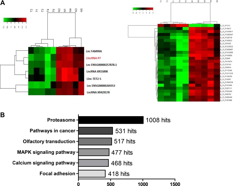
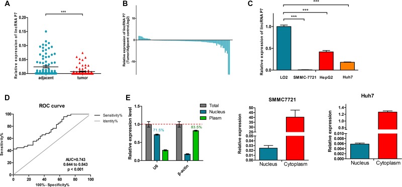
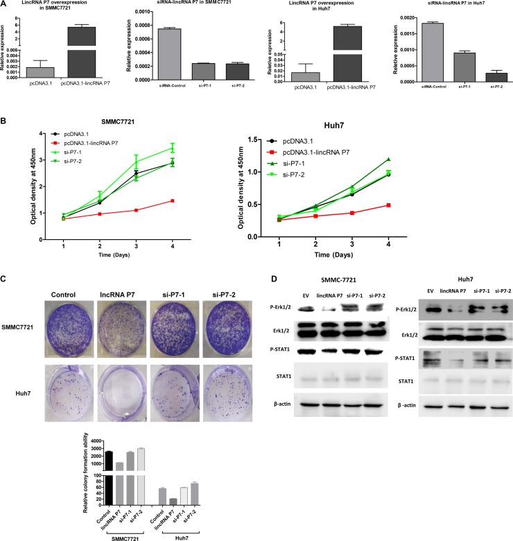
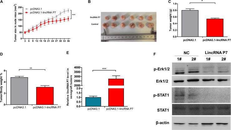
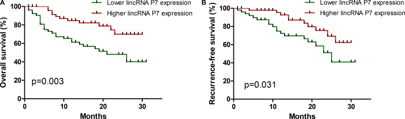
Hepatocellular carcinoma (HCC) is the most common neoplasm and is a leading cause of cancer-related death. Despite advances in the diagnosis and management of HCC, its prognosis remain unfavorable. Accumulating evidence has shown that long intergenic noncoding RNAs (lincRNAs) play central roles in the development of HCC. In this study, we identified a long intergenic noncoding RNA referred to as lincRNA P7 in HCC and explored its clinical significance and biological functions in HCC. The expression level of lincRNA P7 was significantly aberrantly deceased in HCC cancer tissues and cells lines. Gain- and loss-of-function experiments revealed that overexpression of lincRNA P7 significantly inhibited the proliferation of HCC-derived cancer cells, whereas lincRNA P7 knockdown promoted cell growth. Mechanistically, lincRNA P7 blocked Erk1/2 signaling and repressed activation of the STAT1 pathway. In nude mouse models, we show that overexpression of lincRNA P7 effectively repressed HCC xenograft tumor growth in vivo. Moreover, a clinical investigation demonstrated that down-regulated lincRNA P7 expression correlated with liver cirrhosis, Hepatitis B virus (HBV) infection, clinical stage of the tumor and recurrence. A Kaplan-Meier survival analysis showed that the expression of lincRNA P7 was significantly related to overall survival (P = 0.003) and recurrence-free survival (P = 0.031). Collectively, our findings suggested that the down-regulation of lincRNA P7 predicts poor clinical outcomes for HCC patients and might be a powerful candidate prognostic biomarker and target in HCC.
Keywords: HCC; lincRNA P7; long noncoding RNA.
Conflict of interest statement
CONFLICTS OF INTEREST All authors declare no financial or commercial conflicts of interest.
Figures
Similar articles
-
The long intergenic noncoding RNA UFC1, a target of MicroRNA 34a, interacts with the mRNA stabilizing protein HuR to increase levels of β-catenin in HCC cells.Gastroenterology. 2015 Feb;148(2):415-26.e18. doi: 10.1053/j.gastro.2014.10.012. Epub 2014 Oct 22. Gastroenterology. 2015. PMID: 25449213
-
Long noncoding RNA SNHG1 predicts a poor prognosis and promotes hepatocellular carcinoma tumorigenesis.Biomed Pharmacother. 2016 May;80:73-79. doi: 10.1016/j.biopha.2016.02.036. Epub 2016 Mar 15. Biomed Pharmacother. 2016. PMID: 27133041
-
Long noncoding RNA CCHE1 indicates a poor prognosis of hepatocellular carcinoma and promotes carcinogenesis via activation of the ERK/MAPK pathway.Biomed Pharmacother. 2016 Oct;83:450-455. doi: 10.1016/j.biopha.2016.06.056. Epub 2016 Jul 16. Biomed Pharmacother. 2016. PMID: 27427851
-
Long noncoding RNAs: Novel insights into hepatocelluar carcinoma.Cancer Lett. 2014 Mar 1;344(1):20-27. doi: 10.1016/j.canlet.2013.10.021. Epub 2013 Oct 30. Cancer Lett. 2014. PMID: 24183851 Review.
-
An update on long intergenic noncoding RNA p21: a regulatory molecule with various significant functions in cancer.Cell Biosci. 2020 Jun 22;10:82. doi: 10.1186/s13578-020-00445-9. eCollection 2020. Cell Biosci. 2020. PMID: 32582435 Free PMC article. Review.
Cited by
-
Incidence and survival of inflammatory breast cancer between 1973 and 2015 in the SEER database.Breast Cancer Res Treat. 2021 Jan;185(1):229-238. doi: 10.1007/s10549-020-05938-2. Epub 2020 Oct 8. Breast Cancer Res Treat. 2021. PMID: 33033965
-
Potential of miR-181a-5p and miR-630 as clinical biomarkers in NSCLC.BMC Cancer. 2023 Sep 12;23(1):857. doi: 10.1186/s12885-023-11365-5. BMC Cancer. 2023. PMID: 37697308 Free PMC article.
-
Red wavelength-induced photobiomodulation enhances indocyanine green-based anticancer photodynamic therapy.Med Oncol. 2024 Nov 19;42(1):8. doi: 10.1007/s12032-024-02558-4. Med Oncol. 2024. PMID: 39560842
-
Role of FoxP3-positive regulatory T-cells in regressive and progressive cervical dysplasia.J Cancer Res Clin Oncol. 2022 Feb;148(2):377-386. doi: 10.1007/s00432-021-03838-6. Epub 2021 Nov 5. J Cancer Res Clin Oncol. 2022. PMID: 34739585 Free PMC article.
-
Robotic vs laparoscopic approaches of pancreatic resection: a systematic review and meta-analysis.J Robot Surg. 2025 Jun 16;19(1):295. doi: 10.1007/s11701-025-02446-7. J Robot Surg. 2025. PMID: 40522553 Review.
References
-
- Wang H, Dwyer-Lindgren L, Lofgren KT, Rajaratnam JK, Marcus JR, Levin-Rector A, Levitz CE, Lopez AD, Murray CJ. Age-specific and sex-specific mortality in 187 countries, 1970-2010: a systematic analysis for the Global Burden of Disease Study 2010. Lancet. 2012;380:2071–94. doi: 10.1016/S0140-6736(12)61719-X. - DOI - PubMed
LinkOut - more resources
Full Text Sources
Research Materials
Miscellaneous
