Review
. 2018 Nov 15:16:551-564.
doi: 10.1016/j.csbj.2018.11.003. eCollection 2018.

Molecular Mechanisms of Bacterial Bioluminescence

Affiliations
Review

Molecular Mechanisms of Bacterial Bioluminescence

Eveline Brodl et al. Comput Struct Biotechnol J. .

Abstract

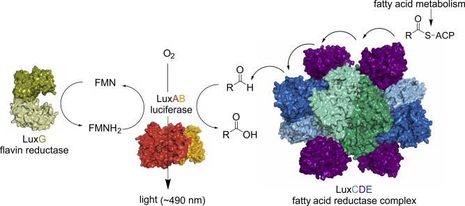
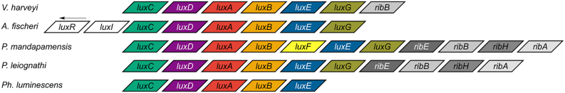
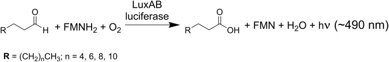
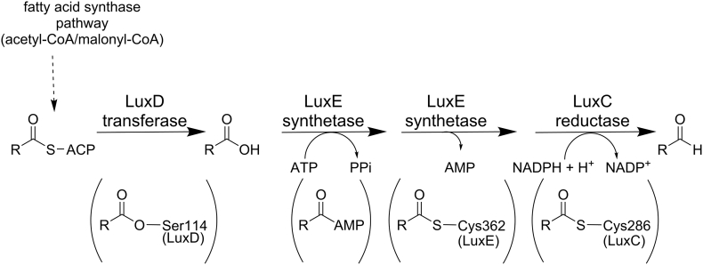
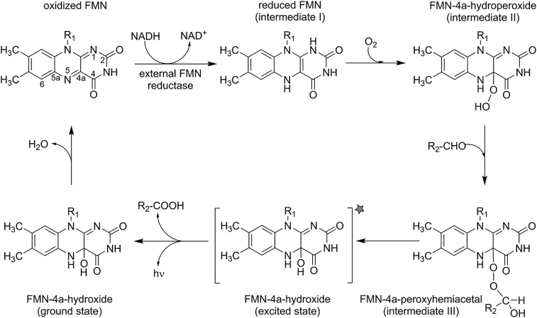
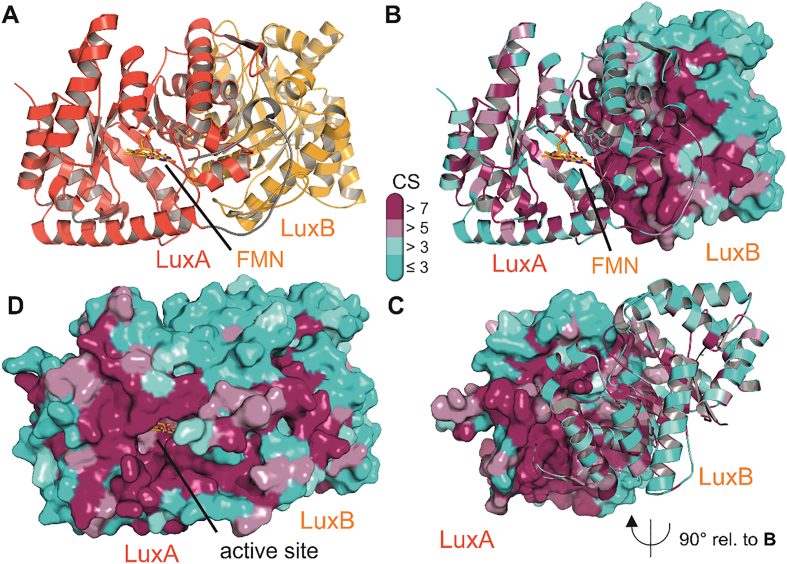
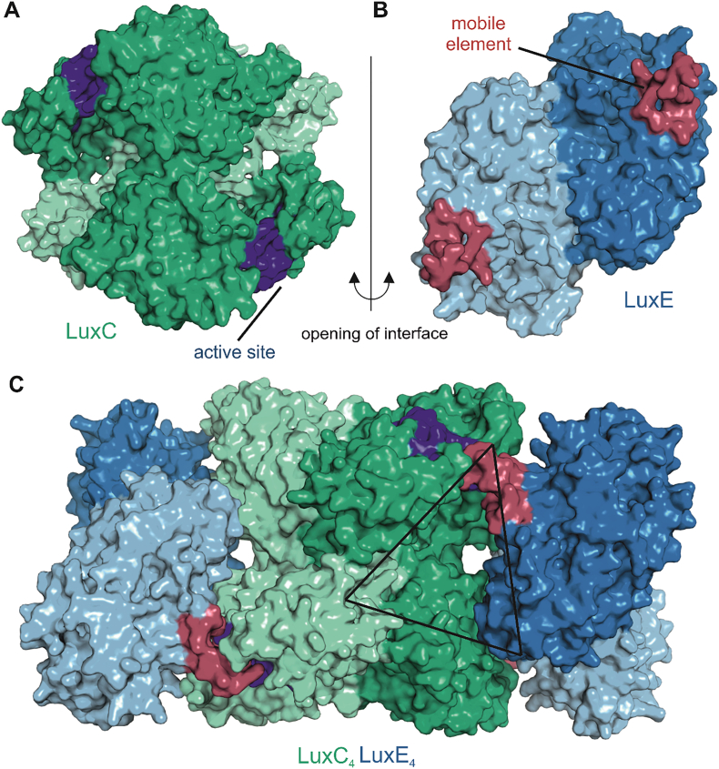
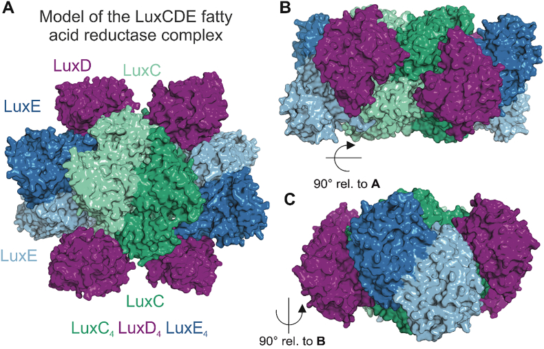
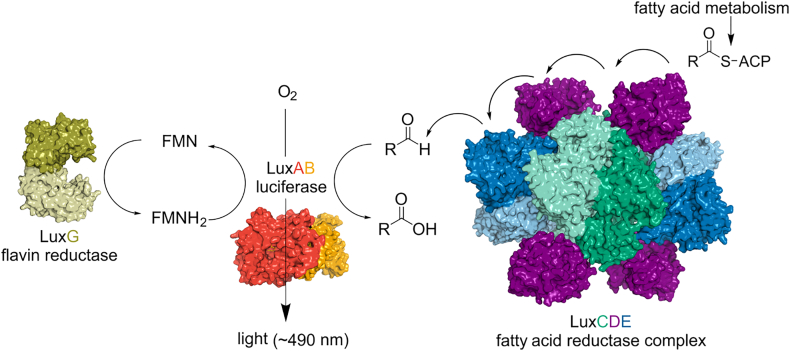
Bioluminescence refers to the production of light by living organisms. Bioluminescent bacteria with a variety of bioluminescence emission characteristics have been identified in Vibrionaceae, Shewanellaceae and Enterobacteriaceae. Bioluminescent bacteria are mainly found in marine habitats and they are either free-floating, sessile or have specialized to live in symbiosis with other marine organisms. On the molecular level, bioluminescence is enabled by a cascade of chemical reactions catalyzed by enzymes encoded by the lux operon with the gene order luxCDABEG. The luxA and luxB genes encode the α- and β- subunits, respectively, of the enzyme luciferase producing the light emitting species. LuxC, luxD and luxE constitute the fatty acid reductase complex, responsible for the synthesis of the long-chain aldehyde substrate and luxG encodes a flavin reductase. In bacteria, the heterodimeric luciferase catalyzes the monooxygenation of long-chain aliphatic aldehydes to the corresponding acids utilizing reduced FMN and molecular oxygen. The energy released as a photon results from an excited state flavin-4a-hydroxide, emitting light centered around 490 nm. Advances in the mechanistic understanding of bacterial bioluminescence have been spurred by the structural characterization of protein encoded by the lux operon. However, the number of available crystal structures is limited to LuxAB (Vibrio harveyi), LuxD (Vibrio harveyi) and LuxF (Photobacterium leiognathi). Based on the crystal structure of LuxD and homology models of LuxC and LuxE, we provide a hypothetical model of the overall structure of the LuxCDE fatty acid reductase complex that is in line with biochemical observations.

Keywords: Bacterial bioluminescence; FMN; Fatty acid reductase complex; Luciferase; Luciferin; Structure-function relationships; lux operon.


Figures

Unlabelled Image
Graphical abstract
Fig. 1
Fig. 1
Examples of lux gene order of bioluminescent bacterial strains (adapted from Fig. 2 in Dunlap P. Bioluminescence: Fundamentals and Applications in Biotechnology - Volume 1. 2014 [6]). According to Table 1 (supplement) and available gene sequences and orders, the five most divergent bacterial strains (Vibrio harveyi, Aliivibrio fischeri, Photobacterium mandapamensis, Photobacterium leiognathi and Photorhabdus luminescens) were chosen to represent lux operon constellations. The color code of individual genes is also used for the corresponding protein models in Fig. 3, Fig. 4, Fig. 5, Fig. 6, Fig. 7, Fig. 8. The arrow above luxR indicates that its reading frame is oriented in the opposite direction (individual operon not directly linked to the lux operon).
Scheme 1
Scheme 1
General reaction mechanism of bacterial bioluminescence. Long-chain aldehydes (CH3(CH2)nCHO), reduced flavin mononucleotide (FMNH2) and molecular oxygen (O2) are converted by the enzyme luciferase (LuxAB) to the corresponding long-chain acids (CH3(CH2)nCOOH), oxidized flavin mononucleotide (FMN), water (H2O) and light emission (hν) with an approximate maximum at 490 nm.
Scheme 2
Scheme 2
Overview of individual reactions catalyzed by the LuxCDE fatty acid reductase complex. Intermediate species covalently linked to individual enzymes are shown in brackets beneath the corresponding reaction steps. The reaction cascade is initiated by the myristoyl transferase LuxD via unloading of myristic acid (R = (CH2)12CH3) bound to the acyl carrier protein (ACP). Covalently linked to Ser114 (Vibrio harveyi numbering) the acyl moiety is transported to the LuxCE complex and released as the free fatty acid that interacts with the LuxE synthetase. At the expense of ATP the fatty acid is activated by LuxE to acyl-AMP and in a second step covalently attached to Cys362. This intermediate is then channeled directly to the active site of the LuxC reductase, where it is initially transferred to Cys286 of LuxC. The latter intermediate is then reduced by NADPH resulting in aldehyde formation and dissociation of the product. Details of the individual processes are provided in the section -The fatty acid reductase complex (luxCDE).
Scheme 3
Scheme 3
Schematic representation of the synthesis pathway of riboflavin (adapted from Fig. 2 in Sung, Lee, J. Photosci., [35]). Guanosine triphosphate (GTP) is converted to 2,5-diamino-6-(5′-phosphoribosylamino)-4-pyrimidineone (DAPO) by RibA, which is further converted in three steps to 5-amino-6-(D-ribitylamino)uracil (APDO). Another route starts from ribulose 5-phosphate, which is converted to 3,4-dihydroxy-2-butanone 4-phosphate (DHBP) by RibB. The enzyme RibH produces the product 6,7-dimethyl-8-ribityllumazine (lumazine) from DHBP and APDO. Lumazine is converted to riboflavin by RibE, which is subsequently transformed into flavin mononucleotide (FMN) by a riboflavin kinase.
Fig. 2
Fig. 2
Bacterial cultures of Photobacterium phosphoreum on the left and Vibrio fischeri Y-1 on the right. The light emission in blue and yellow, respectively, shows nicely the effect of LumP and YFP [47].
Scheme 4
Scheme 4
Reaction cycle of FMN during bacterial bioluminescence. Oxidized flavin mononucleotide (FMN; R1: ribityl monophosphate) is reduced by an external FMN reductase employing NAD(P)H as the reducing agent. Reduced FMN (intermediate I) reacts with dioxygen and forms the FMN-4a-hydroperoxide (intermediate II). The addition of long-chain aldehydes (e.g. R2: (CH2)12CH3) leads to the formation of the FMN-4a-peroxyhemiacetal (intermediate III). After monooxygenation of the long-chain aldehyde to the corresponding acid, an excited state FMN-4a-hydroxide is formed, which is the luciferin in the bacterial bioluminescent reaction. As this luciferin relaxes to the ground state, the free energy is released as light with an emission maximum at 490 nm. After release of one water molecule the catalytic cycle is completed and FMN returned to its oxidized ground state.
Fig. 3
Fig. 3
Crystal structure of the bacterial luciferase from Vibrio harveyi (PDB 3FGC). Panel A shows the characteristic heterodimer of LuxA (red) and LuxB (orange) in cartoon representation. The FMN cofactor in the active site is shown as yellow stick model. The characteristic loop region that mediates the contact between the α- and β-subunits is shown in grey [15]. Panels B, C and D feature the same luciferase dimer colored according to the conservation of residues among the members of bioluminescent bacteria (details of which sequences are aligned are shown in supplementary Table 1). Conservation scores from 1 to 9 correspond to an increase in evolutionary conservation and are colored according to the bar legend in the middle of the figure with higher scores (purple) indicating higher conservation. Panels B and C show the high conservation of residues at the heterodimer interface. Either LuxB or LuxA are shown in surface representation in panels B and C, respectively, and panel C features a 90° out of plane rotation of the dimer for better visibility of the highly conserved LuxA interface. Panel D shows both protomers in surface representation and highlights the strict conservation of residues in the open active site as well as its entrance. Conservation scores were computed with the ConSurf server [11].
Scheme 5
Scheme 5
Mechanistic details of the flavin-initiated electron transfer and the dioxirane mechanism. In the former mechanism (upper reaction path), electron transfer from N5 of the isoalloxazine ring to the distal oxygen atom of the flavin-4a-peroxyhemiacetal (R1: ribityl monophosphate) leads to the formation of a substrate-derived alkoxy radical (e.g. R2: (CH2)12CH3) and the flavin-4a-hydroxy radical cation. Deprotonation of the alkoxy radical generates a resonance stabilized anion radical, which transfers an electron back to the flavin-4a-hydroxy radical cation thus leading to the population of the excited state of the flavin-4a-hydroxide. In an alternative route to this mechanism, the dioxirane mechanism (lower reaction path), the flavin-4a-peroxyhemiacetal forms a dioxirane intermediate, which then receives an electron from the flavin-4a-hydroxide (depicted in dark grey). As before, this leads to the flavin-4a-hydroxide radical cation and the subsequent generation of the excited state similar to the mechanism in the flavin-initiated electron transfer mechanism. In both reactions the rate limiting step is the electron donation from the reduced flavin moiety to the substrate moiety.
Scheme 6
Scheme 6
Proposed mechanism for myrFMN formation (adapted from Scheme 3 in Tabib, Brodl, Macheroux, Mol. Microbiol. 2017 [29]). As shown in Scheme 5, an electron is transferred from the N5 of the flavin to the distal oxygen atom of the peroxy moiety. A hydrogen rearrangement of the alkoxy radical (R2: (CH2)10CH3) intermediate leads to a C3 carbon radical. This combines with the flavin-4a-hydroxide radical cation forming a covalent bond between the C6 of the isoalloxazine ring and the C3 carbon of the myristyl aldehyde. After rearomatization and the oxidation of the aldehyde to the acid followed by release of water, 6-(3′-(R)-myristyl)-FMN (myrFMN) is formed.
Fig. 4
Fig. 4
Crystal structure of LuxD, the myristoyl-ACP-specific thioesterase from V. harveyi (PDB 1THT). Panel A shows the cartoon representation of LuxD in purple color with residues of the catalytic triad as stick models. The flexible hairpin element above the catalytic serine residue is colored in light red. Panels B and C feature the same coloration according to evolutionary conservation of residues introduced in Fig. 3. Highly conserved residues of the whole surface region around the fatty acid binding site support its involvement in complex formation with LuxE and/or LuxC (see below).
Fig. 5
Fig. 5
Homology models of LuxE from V. harveyi. Panels A and B correspond to homology models generated with the SWISS-MODEL server [95] based on PDB 2Y4O and PDB 4R1L, respectively, in the closed and open conformations. The mobile element is depicted in red color and the cysteine residue that can be acylated present in this region is shown as stick model. For reasons of clarity individual functional elements and cofactors are only labelled once in both panels A and B. Nevertheless, all red beta hairpins correspond to the same mobile element and all green stick models correspond to ATP. The overall LuxE dimer is shown in cartoon representation with one protomer colored in blue and the other in light blue. The ATP moiety overlaid from the cofactor bound form of the closed LuxE model template (PDB 2Y27) is shown as green stick model and its proximity to Cys362 further supports the relevance of the closed LuxE state. The open conformation is relevant for the LuxE-LuxC interaction as described before. Panels C and D show the evolutionary conservation of residues according to the ConSurf generated conservation scores (CS – bar legend in the middle). Panel C shows the conservation of the dimer interface in a rotated view relative to panel A and with the light blue protomer shown in transparency. Panel D shows the same view as panels A and B to illustrate the high conservation of residues at a specific surface of the dimer, which is therefore likely involved in the interaction with the LuxC reductase component (cf.Fig. 7) [92].
Fig. 6
Fig. 6
Homology model of LuxC from V. harveyi. Panel A features the tetrameric assembly of LuxC obtained from the SWISS-MODEL server [95] using the crystal structure of methylmalonate semialdehyde dehydrogenase from Bacillus subtilis as template (PDB 1T90). The tetramer corresponds to a dimer of dimers, which are shown once as cartoon representation and once as molecular surface. Individual protomers of each dimer are colored in green and light green. For the dimer in cartoon representation, we also show stick models of the substrate analog (indole-3-acetaldehyde – blue) and the cofactor (NADP+ − grey) in the respective binding sites obtained from the superposition of the LuxC model with indole-3-acetaldehyde dehydrogenase from Pseudomonas syringae (PDB 5IUW) and from the structure of an aldehyde dehydrogenase from Burkholderia multivorans (PDB 5JRY), respectively. Panels B, C and D provide an overview of the evolutionary conservation of residues according to the ConSurf server [11] and computed conservation scores (CS – bar legend). Panel B highlights the conserved residues at the interface of the individual LuxC dimers in the same orientation as panel A. For clarity, the dimer in cartoon representation is shown in transparent mode. Panel C shows a different orientation of the tetrameric assembly, to demonstrate the high degree of conservation in the active site generated at the interface of two subdomains of each LuxC protomer. Importantly, the substrate and the NADPH cofactor approach the active site from opposite sides. Panel D shows another view of one LuxC dimer highlighting the conservation of residues around the NADP+ binding site. For better visibility loop regions covering the NADP+ binding site are not shown in this figure. Similar to the observations for LuxE, patches of strongly conserved residues can be found at surface elements near the active site that are not involved in LuxC oligomerization and are therefore likely involved in complex formation with the LuxE synthetase subunits.
Fig. 7
Fig. 7
Model of the LuxC - LuxE interaction. Panels A and B show surface representations of LuxC and LuxE, respectively, colored according to the details presented in Fig. 5, Fig. 6. The active site region of LuxC is highlighted in dark blue and the mobile element of LuxE containing the acylated residue is colored in red. The central region of complementarity at the LuxC2-LuxE2 interface is lined by many highly conserved residues at specific surfaces of the respective oligomeric structures that have been highlighted in Fig. 5D (LuxE) and Fig. 6B (LuxC). Their complementarity is illustrated by the opening of the interface (right side interface of the complex in panel C) by opposite 90° rotations of LuxC and LuxE. Panel C also shows the second LuxE dimer bound to the opposite side of the LuxC tetramer resulting in an overall LuxC4LuxE4 stoichiometry. The architecture around the active site reveals an interesting triangular complementarity to the LuxD structure (Fig. 4). For generation of the LuxC-LuxE complex we used a different template for homology modeling of LuxC to better reflect the open apo-conformation that might be needed for the initial interaction with LuxE. This model was again generated with the SWISS-MODEL server [95] based on the apo form of the indole-3-acetaldehyde dehydrogenase (PDB 5IUU). The apo form is characterized by a modest opening of the active site accompanied by unstructured loop regions involved in substrate coordination.
Fig. 8
Fig. 8
Tentative model of the LuxCDE fatty acid reductase complex. Panels A, B and C show different views of the LuxC4LuxD4LuxE4 complex. Individual subunits are colored according to their individual presentations in Fig. 4, Fig. 5, Fig. 6. Dark and light colors correspond to individual protomers of the dimeric LuxC and LuxE subspecies. Monomeric LuxD protomers shield off individual active sites of the complex and can readily dissociate [91] to allow release of the final fatty aldehyde product and reassociate to deliver new fatty acid substrates after unloading them from the ACP.
Fig. 9
Fig. 9
Bacterial bioluminescence in a nut shell. The central player in bacterial bioluminescence is the heterodimeric luciferase (red/orange; see Fig. 3), which carries out the oxidation of long-chain fatty aldehydes to the corresponding acid accompanied by light emission (see Scheme 1). The required reduced FMN is provided by a NAD(P)H-dependent FMN reductase (LuxG, on the left side the structure of the closely related enzyme Fre of E. coli is shown in olive; PDB 1QFJ [18]) and the fatty aldehyde is synthesized through the multifunctional complex consisting of LuxCDE (green, violet and blue model on the right; see Fig. 8).

Similar articles

Cited by

References

    1. Widder E.A. Bioluminescence in the ocean: origins of biological, chemical, and ecological diversity. Science. 2010;328:704–708. 80- - PubMed
    1. Dunlap P. In: Bioluminescence, microbial. Schaechter M., editor. Elsevier; Oxford: 2009. pp. 45–61. Encycl Microbiol.
    1. Haddock S.H.D., Moline M.A., Case J.F. Bioluminescence in the sea. Ann Rev Mar Sci. 2010;2:443–493. - PubMed
    1. Hastings J.W., Potrikus C.J., Gupta S.C., Kurfürst M., Makemson J.C. Biochemistry and physiology of bioluminescent bacteria. Adv Microb Physiol. 1985;26:235–291. - PubMed
    1. Zarubin M., Belkin S., Ionescu M., Genin A. Bacterial bioluminescence as a lure for marine zooplankton and fish. Proc Natl Acad Sci. 2012;109:853–857. - PMC - PubMed

LinkOut - more resources