

Measurable Krukenberg tumor is preferably characterized as a non-target lesion in the clinical evaluation of gastric cancer therapeutics: A case report
- PMID: 30546891
- PMCID: PMC6256177
- DOI: 10.3892/mco.2018.1744
Measurable Krukenberg tumor is preferably characterized as a non-target lesion in the clinical evaluation of gastric cancer therapeutics: A case report
Abstract
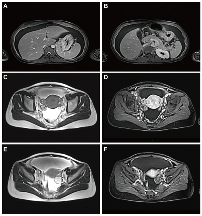
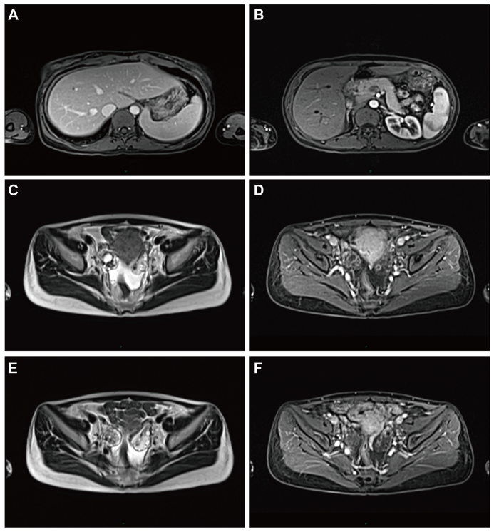
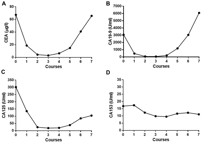
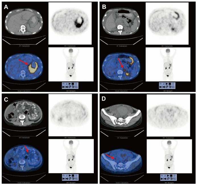
Metastatic cystic lesions may be considered as target lesions according to the Response Evaluation Criteria in Solid Tumors (RECIST) 1.1. However, cystic lesions are considered as non-measurable according to RECIST 1.0. Krukenberg tumors are cystic metastases from gastric cancer. The aim of the present case report was to address the question of whether a Krukenberg tumor can be considered as a target lesion. A 30-year-old female patient was diagnosed with stage IV gastric cancer 6 months after parturition. Subsequently, the patient received two courses of oxaliplatin/capecitabine plus trastuzumab (OCT) treatment. The response evaluation was considered as stable disease. However, after four courses of OCT, the cystic target lesion in the right pelvic cavity exhibited an increase in diameter of >40%. After one more cycle of OCT, contrast-enhanced magnetic resonance imaging (MRI) revealed that the diameter of the cystic mass lesion had decreased by >35% and a further two cycles of treatment were administered. After the last OCT cycle, the levels of the tumor markers cancer antigen (CA) 125, CA19-9 and CA153 had markedly increased, although the cystic mass had decreased in size. Eventually, positron emission tomography-computed tomography (PET/CT) was used to assess the efficacy of treatment. A new lesion was identified, indicating progressive disease. The present case demonstrated that the Krukenberg tumor may be considered as a non-target lesion. In addition, tumor markers and PET/CT yielded results complementary to those of contrast-enhanced MRI in the therapeutic assessment of advanced gastric cancer.
Keywords: Krukenberg tumor; Response Evaluation Criteria in Solid Tumors; gastric cancer; target lesion; therapeutic assessment.
Figures


Similar articles
-
More advantages in detecting bone and soft tissue metastases from prostate cancer using 18F-PSMA PET/CT.Hell J Nucl Med. 2019 Jan-Apr;22(1):6-9. doi: 10.1967/s002449910952. Epub 2019 Mar 7. Hell J Nucl Med. 2019. PMID: 30843003
-
CT and MR findings of Krukenberg tumors: comparison with primary ovarian tumors.J Comput Assist Tomogr. 1996 May-Jun;20(3):393-8. doi: 10.1097/00004728-199605000-00013. J Comput Assist Tomogr. 1996. PMID: 8626898
-
[Rectum-preserving surgery after consolidation neoadjuvant therapy or totally neoadjuvant therapy for low rectal cancer: a preliminary report].Zhonghua Wei Chang Wai Ke Za Zhi. 2020 Mar 25;23(3):281-288. doi: 10.3760/cma.j.cn.441530-20200228-00096. Zhonghua Wei Chang Wai Ke Za Zhi. 2020. PMID: 32192308 Chinese.
-
Diversity of imaging features of ovarian sclerosing stromal tumors on MRI and PET-CT: a case report and literature review.J Ovarian Res. 2018 Dec 20;11(1):101. doi: 10.1186/s13048-018-0473-1. J Ovarian Res. 2018. PMID: 30572921 Free PMC article. Review.
-
Early gastric cancer with Krukenberg tumor and review of cases of intramucosal gastric cancers with Krukenberg tumor.J Gastroenterol. 2003;38(12):1176-80. doi: 10.1007/s00535-003-1227-3. J Gastroenterol. 2003. PMID: 14714257 Review.
References
LinkOut - more resources
Full Text Sources
Research Materials