Review
. 2018 Nov 15:8:47.
doi: 10.4103/jcis.JCIS_45_18. eCollection 2018.

Review of the Major and Minor Salivary Glands, Part 1: Anatomy, Infectious, and Inflammatory Processes

Affiliations
Review

Review of the Major and Minor Salivary Glands, Part 1: Anatomy, Infectious, and Inflammatory Processes

Alexander T Kessler et al. J Clin Imaging Sci. .

Abstract

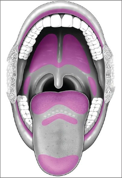
The major and minor salivary glands of the head and neck are important structures that contribute to many of the normal physiologic processes of the aerodigestive tract. The major salivary glands are routinely included within the field of view of standard neuroimaging, and although easily identifiable, salivary pathology is relatively rare and often easy to overlook. Knowledge of the normal and abnormal imaging appearance of the salivary glands is critical for forming useful differential diagnoses, as well as initiating proper clinical workup for what are often incidental findings. The purpose of this review is to provide a succinct image-rich article illustrating relevant anatomy and pathology of the salivary glands via an extensive review of the primary literature. In Part 1, we review anatomy as well as provide an in-depth discussion of the various infectious and inflammatory processes that can affect the salivary glands.

Keywords: Mumps virus; Sjogren's syndrome; salivary gland calculi; salivary glands; sialadenitis.


Conflict of interest statement

There are no conflicts of interest.

Figures

Figure 1
Figure 1
Parotid space anatomy. (a) Illustration of parotid space anatomy with key landmarks labeled. (b) Axial computed tomography of parotid space anatomy with key landmarks labeled. (c) Axial T2-weighted magnetic resonance image of parotid space anatomy with key landmarks labeled.
Figure 2
Figure 2
Parotid (Stensen) duct anatomy. (a) Axial computed tomography of parotid duct anatomy with key landmarks labeled. (b) Axial T2-weighted magnetic resonance image of parotid duct anatomy with key landmarks labeled.
Figure 3
Figure 3
Submandibular space anatomy. (a) Illustration of submandibular and sublingual space anatomy with key landmarks labeled. (b) Axial computed tomography of submandibular space anatomy with key landmarks labeled. (c) Axial T2-weighted magnetic resonance image of submandibular space anatomy with key landmarks labeled. (d) Axial T2-weighted magnetic resonance image of submandibular duct anatomy with key landmarks labeled.
Figure 4
Figure 4
Sublingual space anatomy. (a) Axial computed tomography of sublingual space anatomy with key landmarks labeled. (b) Axial T2-weighted magnetic resonance imaging image of sublingual space anatomy with key landmarks labeled.
Figure 5
Figure 5
Mylohyoid sling/cleft. (a) Axial T2-weighted magnetic resonance image demonstrates the mylohyoid sling with normal free communication posteriorly between submandibular and sublingual spaces (white arrow). (b) Axial T2-weighted magnetic resonance image demonstrates a fat-filled mylohyoid cleft (white arrow). (c) Axial T2-weighted magnetic resonance imaging image demonstrates a mylohyoid cleft containing herniated sublingual gland (white arrow).
Figure 6
Figure 6
Minor salivary gland distribution in the oral cavity (purple).
Figure 7
Figure 7
Sialolithiasis – computed tomography. Axial computed tomography image demonstrates a submandibular duct sialolith (red arrow) with dilated proximal duct (yellow arrow).
Figure 8
Figure 8
Sialolithiasis – Sialography. Right submandibular duct sialogram in a 36-year-old female demonstrates a filling defect (white arrow) at the expected location of the hilum of the submandibular gland, compatible with a calculus.
Figure 9
Figure 9
Viral Sialadenitis. Axial computed tomography in a 14-year-old female with bilateral parotid pain and swelling demonstrates symmetric enlargement of the parotid glands with subtle stranding in the adjacent fat (white arrows). Blood work was positive for parainfluenza virus.
Figure 10
Figure 10
HIV Sialadenitis. A 44-year-old female with HIV not on highly active antiretroviral therapy (CD4 109) presenting with 1 month of progressive painless bilateral parotid swelling. Axial computed tomography demonstrates heterogeneous attenuation throughout the parotid glands with prominent cystic lesions (white arrows), benign lymphoepithelial cysts.
Figure 11
Figure 11
Bacterial sialadenitis. (a) Axial computed tomography in a 70-year-old male with acute right parotid swelling and purulent drainage. (b) Axial computed tomography in a 69-year-old female with acute left submandibular swelling. Both cases demonstrate asymmetric enlargement of a salivary gland with thickening of the adjacent platysma muscle and stranding in the subcutaneous fat (white arrows). Both cases resolved clinically after antibiotics.
Figure 12
Figure 12
Salivary gland abscess. A 69-year-old male presents with left submandibular swelling and purulent drainage. Axial computed tomography demonstrates asymmetric enlargement of the left submandibular gland with thickening of the adjacent platysma and fat stranding. A large 1.2 cm sialolith (white arrow) and 2 cm abscess (yellow arrow) are also present. The patient ultimately required surgical excision of the submandibular gland and abscess drainage.
Figure 13
Figure 13
Early Sjogren's syndrome. Axial computed tomography (a) and axial T2-weighted magnetic resonance image (b) demonstrate enlarged parotid glands with areas of cystic changes (white arrows).
Figure 14
Figure 14
Chronic Sjogren's syndrome. (a) Axial computed tomography demonstrates atrophic parotid glands with multiple parenchymal calcifications. (b) Sialogram of the left submandibular duct demonstrates multifocal areas of narrowing/irregularity through the main duct with pruning of the intraglandular ducts (white arrows).
Figure 15
Figure 15
Sjogren's syndrome with lymphoma. A 76-year-old female with a history of Sjogren's syndrome with bilateral palpable cervical lymph nodes. Axial computed tomography demonstrates multiple mildly enlarged level Ib and IIb lymph nodes (white arrows). Biopsy proved non-Hodgkin lymphoma.
Figure 16
Figure 16
Sarcoid sialadenitis. A 51-year-old male with a history of pulmonary sarcoidosis presents with bilateral parotid swelling. Coronal T1 FS + C images demonstrate marked enlargement and hyperenhancement of the bilateral parotid glands (white arrows), presumably reflecting sarcoid sialadenitis.
Figure 17
Figure 17
Radiation-induced sialadenitis. Axial computed tomography demonstrates hyperenhancement of the bilateral submandibular glands in a patient recently treated with external beam radiation for oropharyngeal cancer.

Similar articles

Cited by

References

    1. Som PM, Brandwin-Gensler MS. Anatomy and pathology of the salivary glands. In: Som PM, Curtin HD, editors. Head and Neck Imaging. 5th ed. St. Louis: Mosby; 2011. pp. 2449–602.
    1. Grossman RI, Yousem DM, editors. Extramucosal diseases of the head and neck. 3rd ed. St. Louis: Mosby; 2010. Neuroradiology: The Requisites; pp. 476–514.
    1. Rastogi R, Bhargava S, Mallarajapatna GJ, Singh SK. Pictorial essay: Salivary gland imaging. Indian J Radiol Imaging. 2012;22:325–33. - PMC - PubMed
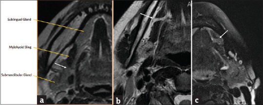
    1. La’porte SJ, Juttla JK, Lingam RK. Imaging the floor of the mouth and the sublingual space. Radiographics. 2011;31:1215–30. - PubMed
    1. Otonari-Yamamoto M, Nakajima K, Tsuji Y, Otonari T, Curtin HD, Okano T, et al. Imaging of the mylohyoid muscle: Separation of submandibular and sublingual spaces. AJR Am J Roentgenol. 2010;194:W431–8. - PubMed