. 2018 Nov 2;8(1):e1523097.
doi: 10.1080/2162402X.2018.1523097. eCollection 2019.

Priming anti-tumor immunity by radiotherapy: Dying tumor cell-derived DAMPs trigger endothelial cell activation and recruitment of myeloid cells

Affiliations

Priming anti-tumor immunity by radiotherapy: Dying tumor cell-derived DAMPs trigger endothelial cell activation and recruitment of myeloid cells

Julia Krombach et al. Oncoimmunology. .

Abstract

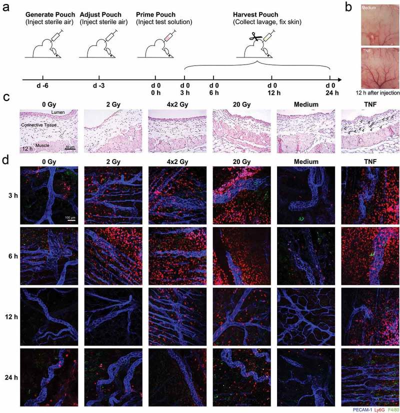
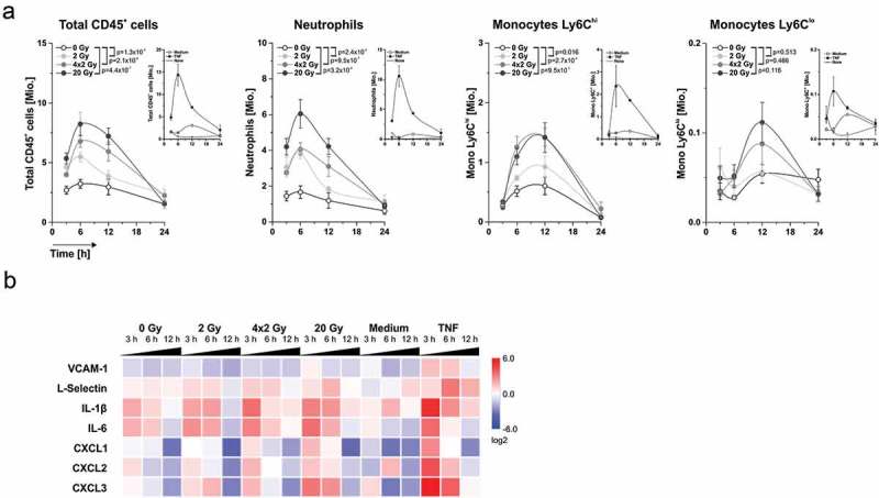
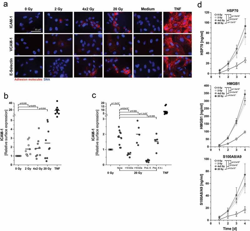
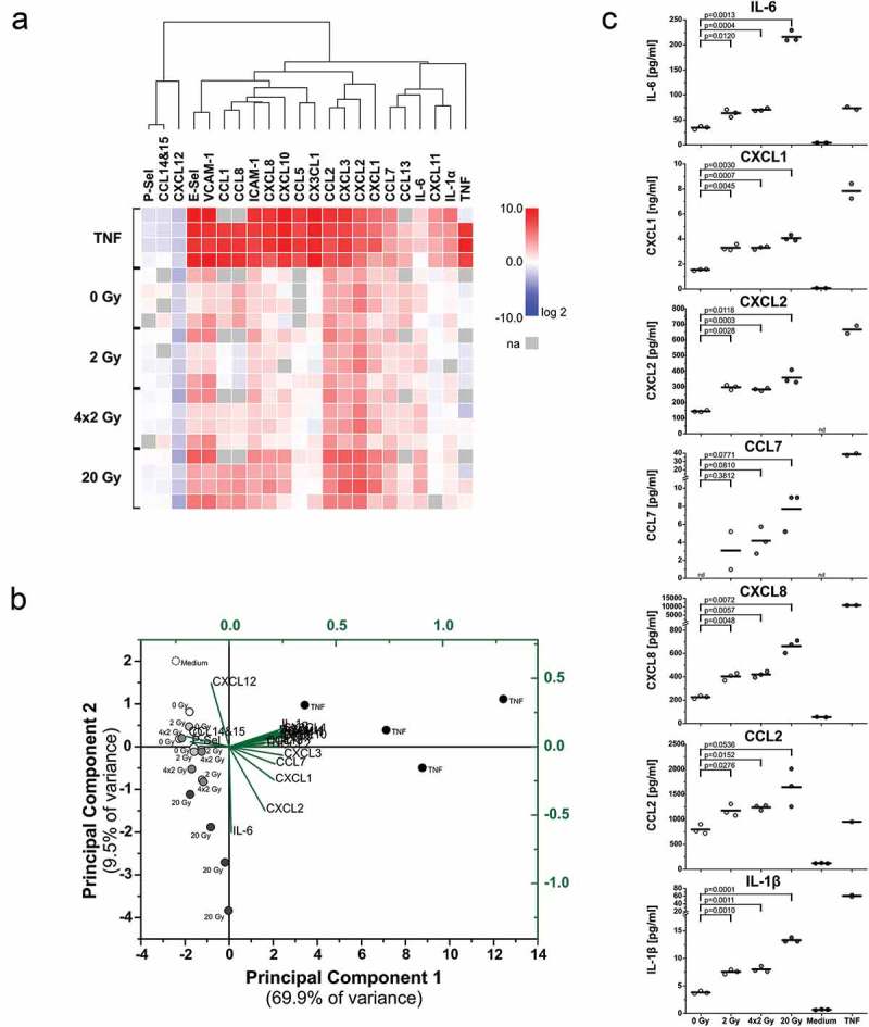
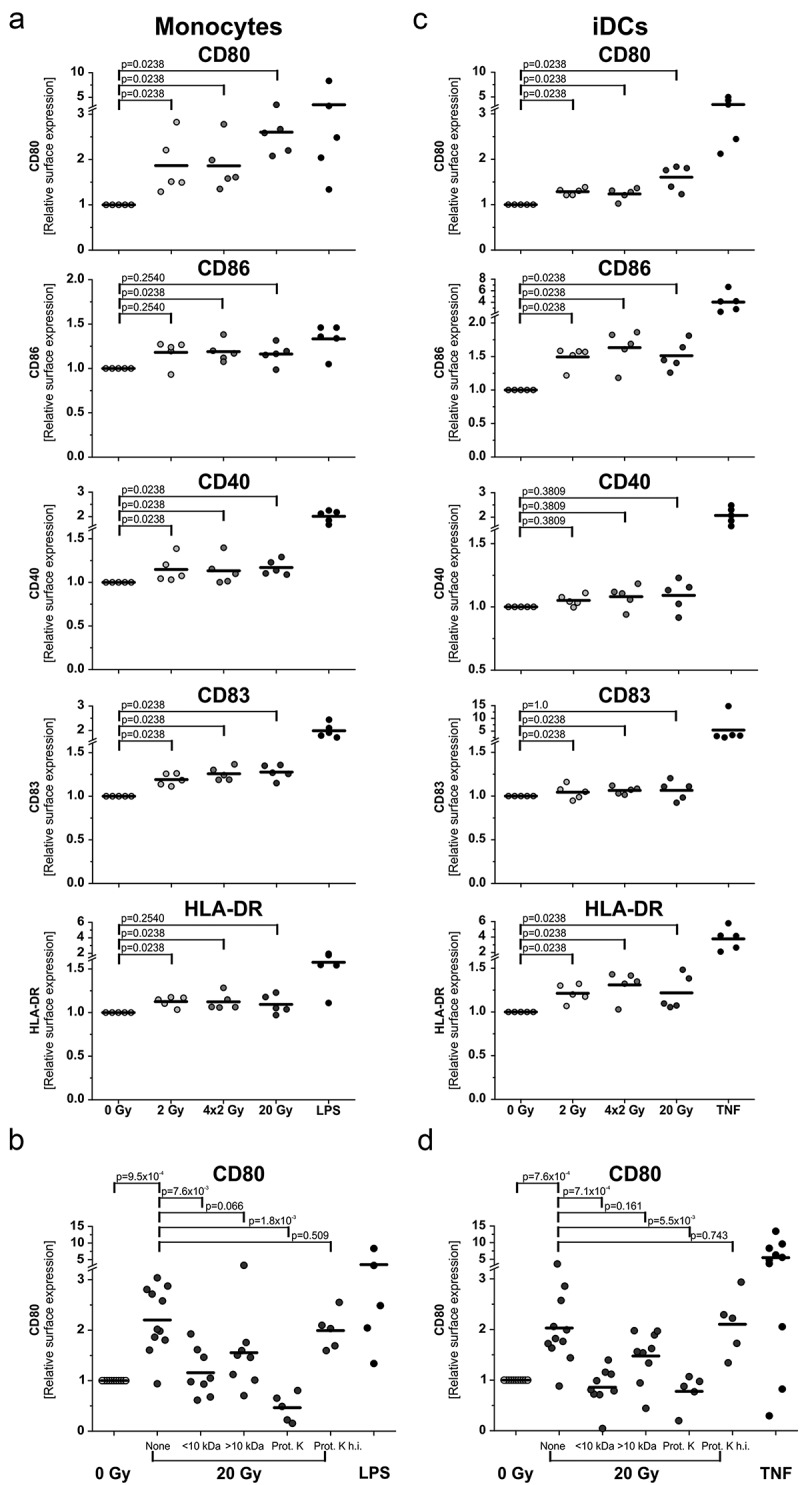
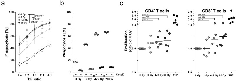
The major goal of radiotherapy is the induction of tumor cell death. Additionally, radiotherapy can function as in situ cancer vaccination by exposing tumor antigens and providing adjuvants for anti-tumor immune priming. In this regard, the mode of tumor cell death and the repertoire of released damage-associated molecular patterns (DAMPs) are crucial. However, optimal dosing and fractionation of radiotherapy remain controversial. Here, we examined the initial steps of anti-tumor immune priming by different radiation regimens (20 Gy, 4 × 2 Gy, 2 Gy, 0 Gy) with cell lines of triple-negative breast cancer in vitro and in vivo. Previously, we have shown that especially high single doses (20 Gy) induce a delayed type of primary necrosis with characteristics of mitotic catastrophe and plasma membrane disintegration. Now, we provide evidence that protein DAMPs released by these dying cells stimulate sequential recruitment of neutrophils and monocytes in vivo. Key players in this regard appear to be endothelial cells revealing a distinct state of activation upon exposure to supernatants of irradiated tumor cells as characterized by high surface expression of adhesion molecules and production of a discrete cytokine/chemokine pattern. Furthermore, irradiated tumor cell-derived protein DAMPs enforced differentiation and maturation of dendritic cells as hallmarked by upregulation of co-stimulatory molecules and improved T cell-priming. Consistently, a recurring pattern was observed: The strongest effects were detected with 20 Gy-irradiated cells. Obviously, the initial steps of radiotherapy-induced anti-tumor immune priming are preferentially triggered by high single doses - at least in models of triple-negative breast cancer.

Keywords: APC recruitment; DAMPs; Radiotherapy; abscopal effects of radiotherapy; anti-tumor immunity; cancer immunotherapy; endothelial cell activation.


Figures

Figure 1.
Figure 1.
In vivo recruitment of myeloid cell subsets stimulated by supernatants of irradiated tumor cells in an air pouch model. (a) Schematic representation of the treatment sequence in air pouch experiments. (b) Representative photographs of native air pouch skin samples (luminal side) 12 h after injection of control medium or medium supplemented with 50 ng/ml TNF. (c) Paraffin sections (3 µm) of representative air pouch skin samples 12 h after injection of the indicated supernatants of irradiated HCC1937 cells or respective control stimuli (medium or 50 ng/ml TNF) were subjected to H&E-staining. 20x magnification, scale bar 50 µm. Arrowheads indicate PMNs in the TNF sample. (d) Exemplary air pouch skin samples were prepared, stained, and examined by confocal immunofluorescence microscopy. For the visualization of endothelial cells, neutrophils, and macrophages, immunostaining against PECAM-1 (blue), Ly6G (red), and F4/80 (green) was performed. 20x magnification, scale bar 100 µm.
Figure 2.
Figure 2.
Dynamics of leukocyte recruitment and mRNA expression of crucial regulator molecules in the air pouch microenvironment. (a) Dynamics of leukocyte recruitment into the air pouch lumen. Air pouch lavages were collected at the indicated time points, and leukocyte subsets were analyzed by flow cytometry. Total cell numbers per pouch are shown (n = 6 animals for supernatants, n = 2 animals for the control stimuli medium or 50 ng/ml TNF). Means ± SEM are depicted, and p-values were calculated by two-way ANOVA with Bonferroni-Holm correction. (b) mRNA expression of key regulator molecules in the air pouch microenvironment. Air pouch skin samples (n = 6 animals for cell culture supernatants, n = 2 animals for the control stimuli medium or 50 ng/ml TNF) were subjected to realtime qRT-PCR. Results were normalized on a reference gene matrix of 18S rRNA, δ-ALAS, β-actin, α-tubulin, and PECAM-1, and calibrated on the medium-injected controls. Means of log2 expression values are depicted.
Figure 3.
Figure 3.
In vitro endothelial cell activation and upregulation of adhesion molecule surface expression are mediated by protein DAMPs derived from irradiated tumor cells. (a) Representative photographs of immunofluorescent adhesion molecule surface staining on HUVECs 4 h after exposure to supernatants of irradiated HCC1937 cells. Surface expression was visualized by immunofluorescence microscopy on native, non-fixed HUVECs. Medium and TNF (50 ng/ml) served as controls. 63x magnification, scale bar 50 µm. (b) Quantitation of ICAM-1 surface expression on HUVECs by fluorometric measurement. HUVECs were treated as in (a) and subjected to native immunofluorescence staining. Staining intensities were quantified by fluorometric measurement, and x-fold expression levels were calculated as the means of fluorescence intensities subtracted by the corresponding isotype controls and normalized to the 0 Gy samples (n = 9 independent experiments). p-values were calculated by unpaired Student’s t-tests with Bonferroni-Holm correction. (c) Biochemical characterization of the molecular entities mediating upregulation of ICAM-1 expression. Supernatants of 20 Gy-irradiated HCC1937 cells were applied to membrane centrifugation (molecular weight cut-off 10 kDa) or proteinase K treatment prior to incubation with HUVECs. ICAM-1 surface expression was measured as in (b) (n = 5–10 independent experiments). Group comparison was performed by unpaired Student’s t-test with Bonferroni-Holm correction. (d) HSP70, HMGB1, and S100A8/A9 were quantified in supernatants of irradiated HCC1937 cells by ELISA. Concentrations were calculated on the basis of standard curves. Means ± SD of 3 (HSP70), 4 (HMGB1), or 5 (S100A8/A9) independent experiments are shown. Group comparison was carried out by two-way ANOVA with Bonferroni-Holm correction.
Figure 4.
Figure 4.
Upregulation of cytokines and chemokines in endothelial cells upon exposure to supernatants of irradiated tumor cells. (a) mRNA expression levels of crucial adhesion molecules, cytokines, and chemokines. HUVECs were incubated with supernatants of irradiated HCC1937 cells for 4 h as in Figure 3. mRNA expression levels were determined by qRT-PCR, normalized on a reference gene matrix of 18S rRNA, β2-microglobulin, and δ-ALAS, and calibrated on the respective medium-treated samples. TNF (50 ng/ml) served as positive control. Unsupervised hierarchical clustering of log2 expression values of 4 independent experiments is depicted (na indicates not assessed). (b) Principal component analysis (PCA) of mRNA expression data from (a). The biplot shows the scores of the samples (black coordinate system) and the scaled loadings of the input variables (green coordinate system) in the subspace of the first two principal components. (c) Cytokines and chemokines released from HUVECs were measured by multiplex-ELISAs after exposure to supernatants of irradiated HCC1937 cells for 4 h and incubation in fresh medium for two more hours. Concentrations were calculated on the basis of standard curves. n = 3 independent experiments are shown (nd indicates not detectable), and p-values were determined by unpaired Student’s t-tests with Bonferroni-Holm correction.
Figure 5.
Figure 5.
Differentiation and maturation of antigen presenting cells is stimulated by protein DAMPs released from irradiated tumor cells. (a) Differentiation of monocyte-derived DCs. Primary human monocytes were stimulated for 4 h with supernatants of irradiated HCC1937 cells followed by differentiation into DCs with 40 ng/ml IL-4 and 20 ng/ml GM-CSF for 5 days. Surface marker expression was measured by flow cytometry. LPS (200 ng/ml) served as positive control. x-fold increase in surface marker expression was calculated from isotype-subtracted median fluorescence intensities normalized on the corresponding 0 Gy samples. Results from 5 independent experiments are shown, and group comparison was performed by two-sided exact Wilcoxon rank test with Bonferroni-Holm correction. (b) Biochemical characterization of the responsible molecular entities. Supernatants of 20 Gy-irradiated HCC1937 cells were subjected to membrane centrifugation (molecular weight cut-off 10 kDa) and proteinase K digestion prior to incubation with monocytes. CD80 surface expression was determined as in (a). Data from 5–10 independent experiments are shown, and group comparison was performed by two-sided exact Wilcoxon rank test with Bonferroni-Holm correction. (c) Maturation of immature DCs. Immature DCs were differentiated from primary human monocytes with IL-4 (40 ng/ml) and GM-CSF (20 ng/ml) for 5 days. DCs were then stimulated with supernatants of irradiated HCC1937 cells for 2 days and examined by flow cytometry. TNF (100 ng/ml) served as positive control. Data from 5 independent experiments are presented, and p-values were calculated by two-sided exact Wilcoxon rank test with Bonferroni-Holm correction. (d) Biochemical characterization. Prior to incubation with DCs, supernatants of 20 Gy-irradiated HCC1937 cells were applied to membrane centrifugation or proteinase K digestion, respectively, as in (b). Data from 5–10 independent experiments are shown, and group comparison was performed by two-sided exact Wilcoxon rank test with Bonferroni-Holm correction.
Figure 6.
Figure 6.
Effector functions of antigen presenting cells are enhanced upon contact with irradiated tumor cells. (a) Phagocytosis and antigen uptake. Immature DCs were differentiated from primary human monocytes (PKH67-labeled) with 40 ng/mL IL-4 and 20 ng/mL GM-CSF for 5 days. Afterwards, DCs were co-incubated with irradiated HCC1937 cells (4 days after irradiation, PKH26-labeled) at the indicated target:effector ratios. Phagocytosis was allowed for 2 h and analyzed by flow cytometry. The percentage of double-positive DCs with ingested HCC1937 cell material is shown as means ± SD of 5 independent experiments. Group comparison was calculated by two-way ANOVA with Bonferroni-Holm correction. (b) DCs were incubated with 20 µM cytochalasin D 1 h prior to performing the phagocytosis assay at a ratio of 1:4 as in (a). (c) Priming of T cell proliferation. DCs were differentiated from primary human monocytes upon exposure to supernatants of irradiated HCC1937 cells or TNF (100 ng/ml) as in Figure 5A. After 7 days, DCs were co-incubated with CFSE-labeled T cells from an allogeneic donor at a ratio of 1:5 (DC:T cells) for additional 5 days before T cell proliferation was analyzed by flow cytometry. The percentage of proliferating T cells was calculated as the percentage of CD3+CFSElowCD4+ or CD3+CFSElowCD8+ on the basis of all CD3+CD4+ or CD3+CD8+ cells, respectively. Results were normalized on the corresponding 0 Gy samples and are displayed as data from 6 independent experiments. Group comparison was carried out by two-sided exact Wilcoxon rank test with Bonferroni-Holm correction.

Similar articles

Cited by

References

    1. Orth M, Lauber K, Niyazi M, Friedl AA, Li M, Maihofer C, Schuttrumpf L, Ernst A, Niemoller OM, Belka C.. Current concepts in clinical radiation oncology. Radiat Environ Biophys. 2014;53:1–29.doi:10.1007/s00411-013-0497-2. - PMC - PubMed
    1. Garg AD, Romano E, Rufo N, Agostinis P. Immunogenic versus tolerogenic phagocytosis during anticancer therapy: mechanisms and clinical translation. Cell Death Differ. 2016;23:938–951. doi:10.1038/cdd.2016.5. - DOI - PMC - PubMed
    1. Lauber K, Ernst A, Orth M, Herrmann M, Belka C. Dying cell clearance and its impact on the outcome of tumor radiotherapy. Front Oncol. 2012;2:116. doi:10.3389/fonc.2012.00116. - DOI - PMC - PubMed
    1. Demaria S, Formenti SC. Radiation as an immunological adjuvant: current evidence on dose and fractionation. Front Oncol. 2012;2:153. doi:10.3389/fonc.2012.00153. - DOI - PMC - PubMed
    1. Chen DS, Mellman I. Oncology meets immunology: the cancer-immunity cycle. Immunity. 2013;39:1–10. doi:10.1016/j.immuni.2013.07.012. - DOI - PubMed

Publication types

LinkOut - more resources